


Introduction
Malignant melanoma is the leading cause of cancer-related deaths worldwide, and its incidence in China has been increasing in the recent years [1, 2]. The treatment of advanced melanoma has remarkably improved due to the successful clinical development of immune checkpoint inhibitors (ICIs) that reactivate the anticancer immune response [3, 4]. The anti-programmed cell death 1 (PD-1) receptor antibody represents the second breakthrough in immune checkpoint blockade therapy of melanoma after the approval of ipilimumab [5–8]. Two anti-PD-1 antibodies, pembrolizumab, and nivolumab were approved for the treatment of melanoma in 2014 [6, 9].
PD-1 is a key inhibitory receptor expressed on activated CD4+ T cells, CD8+ T cells, natural killer T cells, and B cells [10–14]. Its binding with programmed cell death ligands (PD-L1 and PD-L2) expressed on antigen-presenting cells, and human cancers including melanoma delivers a negative signal to lymphocytes that inhibits T-cell proliferation, cytokine release, and cytotoxicity [10–14]. Anti-PD-1 antibodies including pembrolizumab and nivolumab can reverse this T-cell suppression and induce long-lasting antitumor responses in patients with advanced solid tumors, including advanced melanoma [15–17]. By activating the immune system, the anti-PD-1 antibodies lead to autoimmune-like toxicities known as immune-related adverse events (irAEs) through immune cell infiltration into normal, noncancerous tissues [6]. Such irAEs were not frequently observed with cytotoxic chemotherapy or other classes of targeted agents [5, 18]. The irAEs in response to ICIs have varying times to onset and include organ-specific toxicities in skin, endocrine, gastrointestinal, hepatobiliary, pulmonary, and renal, as well as non-organ-specific toxicities such as fatigue, pyrexia, appetite loss, arthralgia, and myalgia [19–22]. Their presentation can range from mild and manageable, to severe and life threatening if not recognized early and treated with appropriate measures such as corticosteroids [5, 23].
Recent studies have demonstrated that the development of irAEs in melanoma [11, 24–27] and non–small cell lung cancer (NSCLC) [12, 18, 20, 28, 29] patients treated with ICIs could correlate with the clinical response. Chinese melanoma patients have a higher proportion of the acral and mucosal types, which have distinct genetic and clinical characteristics, lower somatic mutational burden, and poorer prognoses [30–33]. However, the relationship between the irAEs and clinical outcomes remains unclear. In this study, we performed a retrospective analysis of the clinical data obtained from 93 advanced melanoma patients treated with anti-PD-1 antibodies at the Sun Yat-sen University Cancer Center and evaluated the association between irAEs and clinical outcomes. The results of this study will help identify patients with advanced melanoma who are most likely to benefit from PD-1 checkpoint inhibitor therapy.
Results
Patient characteristics
As list in Table 1, a total of 93 patients with advanced melanoma treated with PD-1 inhibitors at our center between August 2014 and March 2018 were enrolled in this study. Of these patients, 59 (63.4%) and 34 (36.6%) were treated with pembrolizumab and nivolumab, respectively. The median number of doses of anti-PD-1 inhibitors doses was 5 (range, 2–24). The cohort comprised of 54 men (58.1%) and 39 women (41.9%), with a median age of 52 years (range, 22-78 years). The primary lesions were acral melanomas that arose from palms, soles, and subungual sites in 26 patients (28.0%), chronic sun-derived (CSD) or non-CSD melanomas that arose in non-acral sites in 34 patients (36.5%), mucosal melanomas in 21 patients (22.6%) and uveal in 3 patients (3.2%). Nine patients (9.7%) had no primary lesions. While 33 patients (35.5%) had elevated serum LDH levels, 21 (22.6%) harbored BRAF V600E mutation and 2 (2.2%) harbored C-KIT mutation. Ten patients (10.8%) had brain metastases, and they received radiotherapy for the brain lesions before the infusion of anti-PD-1 antibodies. A total of 61 patients (65.6%) had received prior ipilimumab, chemotherapy or BRAF inhibitors. Overall, irAEs were noted in 54 patients (58.1%). No significant differences were observed in the baseline characteristics of the patients with and without irAEs (Table 1, P > 0.05).
Table 1. Distribution of demographic and clinical characteristics of patients.
| Characteristics | Patients no. (%) | ||||||||||||||||||||||||||||||||||||||||||||||||||||||||||||||||||||||||||||||||||||||||||||||||||
| Total (n=93) | With irAEs (n=54) | Without irAEs (n=39) | P | ||||||||||||||||||||||||||||||||||||||||||||||||||||||||||||||||||||||||||||||||||||||||||||||||
| Age, mean(range) | 52(22-78) | 52(25-77) | 53(22-78) | 0.827 | |||||||||||||||||||||||||||||||||||||||||||||||||||||||||||||||||||||||||||||||||||||||||||||||
| Gender | 0.784 | ||||||||||||||||||||||||||||||||||||||||||||||||||||||||||||||||||||||||||||||||||||||||||||||||||
| male | 54(58.1) | 32(59.3) | 22(56.4) | ||||||||||||||||||||||||||||||||||||||||||||||||||||||||||||||||||||||||||||||||||||||||||||||||
| female | 39(41.9) | 22(40.7) | 17(43.6) | ||||||||||||||||||||||||||||||||||||||||||||||||||||||||||||||||||||||||||||||||||||||||||||||||
| ECOG status | 0.207 | ||||||||||||||||||||||||||||||||||||||||||||||||||||||||||||||||||||||||||||||||||||||||||||||||||
| 0-1 | 84(90.3) | 47(87.0) | 37(94.9) | ||||||||||||||||||||||||||||||||||||||||||||||||||||||||||||||||||||||||||||||||||||||||||||||||
| ≥2 | 9(9.7) | 7(13.0) | 2(5.1) | ||||||||||||||||||||||||||||||||||||||||||||||||||||||||||||||||||||||||||||||||||||||||||||||||
| Primary site | 0.653 | ||||||||||||||||||||||||||||||||||||||||||||||||||||||||||||||||||||||||||||||||||||||||||||||||||
| Acral | 26(28.0) | 18(33.3) | 8(20.5) | ||||||||||||||||||||||||||||||||||||||||||||||||||||||||||||||||||||||||||||||||||||||||||||||||
| CSD/non-CSD | 34(36.5) | 19(35.2) | 15(38.5) | ||||||||||||||||||||||||||||||||||||||||||||||||||||||||||||||||||||||||||||||||||||||||||||||||
| Mucosal | 21(22.6) | 10(18.5) | 11(28.2) | ||||||||||||||||||||||||||||||||||||||||||||||||||||||||||||||||||||||||||||||||||||||||||||||||
| Uveal | 3(3.2) | 2(3.7) | 1(2.6) | ||||||||||||||||||||||||||||||||||||||||||||||||||||||||||||||||||||||||||||||||||||||||||||||||
| No-primary lesion | 9(9.7) | 5(9.3) | 4(10.2) | ||||||||||||||||||||||||||||||||||||||||||||||||||||||||||||||||||||||||||||||||||||||||||||||||
| Metastasis stage a | 0.719 | ||||||||||||||||||||||||||||||||||||||||||||||||||||||||||||||||||||||||||||||||||||||||||||||||||
| M1a | 24(25.8) | 15(27.8) | 9(23.1) | ||||||||||||||||||||||||||||||||||||||||||||||||||||||||||||||||||||||||||||||||||||||||||||||||
| M1b | 20(21.5) | 13(24.1) | 7(17.9) | ||||||||||||||||||||||||||||||||||||||||||||||||||||||||||||||||||||||||||||||||||||||||||||||||
| M1c | 39(41.9) | 20(37.0) | 19(48.7) | ||||||||||||||||||||||||||||||||||||||||||||||||||||||||||||||||||||||||||||||||||||||||||||||||
| M1d | 10(10.8) | 6(11.1) | 4(10.3) | ||||||||||||||||||||||||||||||||||||||||||||||||||||||||||||||||||||||||||||||||||||||||||||||||
| LDH level | 0.944 | ||||||||||||||||||||||||||||||||||||||||||||||||||||||||||||||||||||||||||||||||||||||||||||||||||
| ≤UNL | 60(64.5) | 35(64.8) | 25(64.1) | ||||||||||||||||||||||||||||||||||||||||||||||||||||||||||||||||||||||||||||||||||||||||||||||||
| >UNL | 33(35.5) | 19(35.2) | 14(35.9) | ||||||||||||||||||||||||||||||||||||||||||||||||||||||||||||||||||||||||||||||||||||||||||||||||
| Brain metastasis | 0.896 | ||||||||||||||||||||||||||||||||||||||||||||||||||||||||||||||||||||||||||||||||||||||||||||||||||
| Yes | 10(10.8) | 6(11.1) | 4(10.3) | ||||||||||||||||||||||||||||||||||||||||||||||||||||||||||||||||||||||||||||||||||||||||||||||||
| No | 83(89.2) | 48(88.9) | 35(89.7) | ||||||||||||||||||||||||||||||||||||||||||||||||||||||||||||||||||||||||||||||||||||||||||||||||
| Liver metastasis | 0.484 | ||||||||||||||||||||||||||||||||||||||||||||||||||||||||||||||||||||||||||||||||||||||||||||||||||
| Yes | 32(34.4) | 17(31.5) | 15(38.5) | ||||||||||||||||||||||||||||||||||||||||||||||||||||||||||||||||||||||||||||||||||||||||||||||||
| No | 61(65.6) | 37(68.5) | 24(61.5) | ||||||||||||||||||||||||||||||||||||||||||||||||||||||||||||||||||||||||||||||||||||||||||||||||
| Lung metastasis | 0.978 | ||||||||||||||||||||||||||||||||||||||||||||||||||||||||||||||||||||||||||||||||||||||||||||||||||
| Yes | 55(59.1) | 32(59.3) | 23(59.0) | ||||||||||||||||||||||||||||||||||||||||||||||||||||||||||||||||||||||||||||||||||||||||||||||||
| No | 38(40.9) | 22(40.7) | 16(41.0) | ||||||||||||||||||||||||||||||||||||||||||||||||||||||||||||||||||||||||||||||||||||||||||||||||
| BRAF V600E status | 0.270 | ||||||||||||||||||||||||||||||||||||||||||||||||||||||||||||||||||||||||||||||||||||||||||||||||||
| mutation | 21(22.6) | 10(18.5) | 11(28.2) | ||||||||||||||||||||||||||||||||||||||||||||||||||||||||||||||||||||||||||||||||||||||||||||||||
| wild-type | 72(77.4) | 44(81.5) | 28(71.8) | ||||||||||||||||||||||||||||||||||||||||||||||||||||||||||||||||||||||||||||||||||||||||||||||||
| C-KIT status | 0.093 | ||||||||||||||||||||||||||||||||||||||||||||||||||||||||||||||||||||||||||||||||||||||||||||||||||
| mutation | 2 | 0(0) | 2(5.1%) | ||||||||||||||||||||||||||||||||||||||||||||||||||||||||||||||||||||||||||||||||||||||||||||||||
| wild-type | 91 | 54(100%) | 37(94.9%) | ||||||||||||||||||||||||||||||||||||||||||||||||||||||||||||||||||||||||||||||||||||||||||||||||
| Prior therapy b | 0.530 | ||||||||||||||||||||||||||||||||||||||||||||||||||||||||||||||||||||||||||||||||||||||||||||||||||
| Yes | 61(65.6) | 34(63.0) | 27(69.2) | ||||||||||||||||||||||||||||||||||||||||||||||||||||||||||||||||||||||||||||||||||||||||||||||||
| No | 32(34.4) | 20(37.0) | 12(30.8) | ||||||||||||||||||||||||||||||||||||||||||||||||||||||||||||||||||||||||||||||||||||||||||||||||
| PD-1 inhibitor | 0.324 | ||||||||||||||||||||||||||||||||||||||||||||||||||||||||||||||||||||||||||||||||||||||||||||||||||
| Pembrolizumab | 59(63.4) | 32(59.3) | 27(69.2) | ||||||||||||||||||||||||||||||||||||||||||||||||||||||||||||||||||||||||||||||||||||||||||||||||
| Nivolumab | 34(36.6) | 22(40.7) | 12(30.8) | ||||||||||||||||||||||||||||||||||||||||||||||||||||||||||||||||||||||||||||||||||||||||||||||||
| Abbreviations: ECOG, Eastern Cooperative Oncology Group; CSD, chronic sun derived; LDH, Lactate dehydrogenase; ULN, upper limit of normal | |||||||||||||||||||||||||||||||||||||||||||||||||||||||||||||||||||||||||||||||||||||||||||||||||||
| a According to the 7th edition of the AJCC staging manual. | |||||||||||||||||||||||||||||||||||||||||||||||||||||||||||||||||||||||||||||||||||||||||||||||||||
| b Including ipilimumab, chemotherapy or BRAF inhibitors for patients with BRAF mutation. | |||||||||||||||||||||||||||||||||||||||||||||||||||||||||||||||||||||||||||||||||||||||||||||||||||
IrAEs profile of the patients
IrAEs occurred in 58.1% of the patients (54/93), which were grade 1-2 in 46 patients (49.5%) and grade 3-4 in 8 (8.6%) patients (Table 2). The observed irAEs were skin (67.8%), endocrine (11.8%), gastrointestinal (6.5%), hepatobiliary (17.2%), and others (25.8%) (Table 2). No pulmonary and renal events were reported in this study population. The most common skin irAEs were pruritus (30.1%), rash (24.7%), and vitiligo (16.1%), and the most common endocrine irAEs were thyroiditis/hypothyroidism (7.5%) and hypoadrenocorticism (6.5%). Hepatitis (8.6%) and elevated transaminase levels (7.5%) were the most common hepatobiliary irAEs observed (Table 2). The other irAEs included fatigue (15.1%) and pyrexia (8.6%). Steroids were used to treat the irAEs in 8 patients (8.6%) (Table 2). Median time to onset in weeks was 7.4 (range, 0.1-36.3), 11.9 (1.0-36.4), 4.6 (0.4-21.7), 6.6 (1.0-24.7) and 3.9 (0.1-18.6) for skin, endocrine, gastrointestinal, hepatobiliary and other irAEs, respectively, of any grade (Table 2).
Table 2. Immune-related adverse events according to category and grade.
| Category | Patients no. (%) | Weeks to Onset, Median (range) | |||||||||||||||||||||||||||||||||||||||||||||||||||||||||||||||||||||||||||||||||||||||||||||||||
| Total (n=93) | Grade 1-2 | Grade 3-4 | Systemic Steroid Therapy | ||||||||||||||||||||||||||||||||||||||||||||||||||||||||||||||||||||||||||||||||||||||||||||||||
| Any | 54(58.1) | 46(49.5) | 8(8.6) | 8(8.6) | 6.1(0.1-36.4) | ||||||||||||||||||||||||||||||||||||||||||||||||||||||||||||||||||||||||||||||||||||||||||||||
| Skin | 40(67.8) | 7.4(0.1-36.3) | |||||||||||||||||||||||||||||||||||||||||||||||||||||||||||||||||||||||||||||||||||||||||||||||||
| Rash | 23(24.7) | 23(24.7) | NA | NA | |||||||||||||||||||||||||||||||||||||||||||||||||||||||||||||||||||||||||||||||||||||||||||||||
| Pruritus | 28(30.1) | 28(30.1) | NA | NA | |||||||||||||||||||||||||||||||||||||||||||||||||||||||||||||||||||||||||||||||||||||||||||||||
| Vitiligo | 15(16.1) | 15(16.1) | NA | NA | |||||||||||||||||||||||||||||||||||||||||||||||||||||||||||||||||||||||||||||||||||||||||||||||
| Psoriasis | 1(1.1) | 1(1.1) | NA | NA | |||||||||||||||||||||||||||||||||||||||||||||||||||||||||||||||||||||||||||||||||||||||||||||||
| Endocrine | 11(11.8) | 11.9(1.0-36.4) | |||||||||||||||||||||||||||||||||||||||||||||||||||||||||||||||||||||||||||||||||||||||||||||||||
| Thyroiditis/hypothyroidism | 7(7.5) | 7(7.5) | NA | NA | |||||||||||||||||||||||||||||||||||||||||||||||||||||||||||||||||||||||||||||||||||||||||||||||
| Hypophysitis | 1(1.1) | 1(1.1) | NA | NA | |||||||||||||||||||||||||||||||||||||||||||||||||||||||||||||||||||||||||||||||||||||||||||||||
| Hypoadrenocorticism | 6(6.5) | 6(6.5) | NA | NA | |||||||||||||||||||||||||||||||||||||||||||||||||||||||||||||||||||||||||||||||||||||||||||||||
| Gastrointestinal | 6(6.5) | 4.6(0.4-21.7) | |||||||||||||||||||||||||||||||||||||||||||||||||||||||||||||||||||||||||||||||||||||||||||||||||
| Diarrhea | 5(5.4) | 5(5.4) | NA | NA | |||||||||||||||||||||||||||||||||||||||||||||||||||||||||||||||||||||||||||||||||||||||||||||||
| Nausea/vomiting | 1(1.1) | 1(1.1) | NA | NA | |||||||||||||||||||||||||||||||||||||||||||||||||||||||||||||||||||||||||||||||||||||||||||||||
| colitis | NA | NA | NA | NA | |||||||||||||||||||||||||||||||||||||||||||||||||||||||||||||||||||||||||||||||||||||||||||||||
| Hepatobiliary | 16(17.2) | 6.6(1.0-24.7) | |||||||||||||||||||||||||||||||||||||||||||||||||||||||||||||||||||||||||||||||||||||||||||||||||
| Elevated transaminase | 7(7.5) | 7(7.5) | NA | NA | |||||||||||||||||||||||||||||||||||||||||||||||||||||||||||||||||||||||||||||||||||||||||||||||
| Hyperbilirubinemia | 1(1.1) | 1(1.1) | NA | NA | |||||||||||||||||||||||||||||||||||||||||||||||||||||||||||||||||||||||||||||||||||||||||||||||
| Hepatitis | 8(8.6) | 3(3.2) | 5(5.4) | 7(7.5) | |||||||||||||||||||||||||||||||||||||||||||||||||||||||||||||||||||||||||||||||||||||||||||||||
| Cholangitis | NA | NA | NA | NA | |||||||||||||||||||||||||||||||||||||||||||||||||||||||||||||||||||||||||||||||||||||||||||||||
| Pulmonary | NA | NA | NA | NA | |||||||||||||||||||||||||||||||||||||||||||||||||||||||||||||||||||||||||||||||||||||||||||||||
| Renal | NA | NA | NA | NA | |||||||||||||||||||||||||||||||||||||||||||||||||||||||||||||||||||||||||||||||||||||||||||||||
| Other | 24(25.8) | 3.9(0.1-18.6) | |||||||||||||||||||||||||||||||||||||||||||||||||||||||||||||||||||||||||||||||||||||||||||||||||
| Fatigue | 14(15.1) | 14(15.1) | NA | NA | |||||||||||||||||||||||||||||||||||||||||||||||||||||||||||||||||||||||||||||||||||||||||||||||
| Appetite loss | 3(3.2) | 3(3.2) | NA | NA | |||||||||||||||||||||||||||||||||||||||||||||||||||||||||||||||||||||||||||||||||||||||||||||||
| Arthralgia/myalgia | 3(3.2) | 3(3.2) | NA | NA | |||||||||||||||||||||||||||||||||||||||||||||||||||||||||||||||||||||||||||||||||||||||||||||||
| Pyrexia | 8(8.6) | 5(5.4) | 3(3.2) | NA | |||||||||||||||||||||||||||||||||||||||||||||||||||||||||||||||||||||||||||||||||||||||||||||||
| Rhabdomyolysis | 1(1.1) | NA | 1(1.1) | 1(1.1) | |||||||||||||||||||||||||||||||||||||||||||||||||||||||||||||||||||||||||||||||||||||||||||||||
| Uveitis | 1(1.1) | NA | 1(1.1) | 1(1.1) | |||||||||||||||||||||||||||||||||||||||||||||||||||||||||||||||||||||||||||||||||||||||||||||||
| Abbreviations: NA, not applicable. | |||||||||||||||||||||||||||||||||||||||||||||||||||||||||||||||||||||||||||||||||||||||||||||||||||
Association between irAEs and response rates
In this study, the objective response rate (ORR) and disease control rate (DCR) in patients who received PD-1 inhibitors were 22.6% and 40.9%, respectively (Table 3). The ORR and DCR were significantly better in patients who experienced irAEs than those who did not (33.3% versus 7.7%; P = 0.004 and 53.7% versus 23.1%; P = 0.003, respectively) (Table 3). The ORR and DCR were a little higher in patients who experienced one to two irAEs than those with no irAEs (19.4% versus 7.7% and 35.5% versus 23.1%, respectively), although the results were not statistically significant (P = 0.148 and P = 0.254, respectively) (Table 3). Moreover, ORR and DCR were significantly better in patients who experienced three or more irAEs than those who experienced no irAEs (ORR: 42.2% versus 7.7%; P < 0.001 and DCR: 78.3% versus 23.1%; P < 0.001) and one to two irAEs (ORR: 42.2% versus 19.4%; P < 0.001 and DCR: 78.3% versus 35.5% P < 0.001) (Table 3). In addition, patients with grade 1 to 2 irAEs had significantly higher ORR and DCR than those with no irAEs (40.0% versus 7.7%; P = 0.002 and 54.3% versus 23.1%; P = 0.003, respectively) (Table 3). In contrast, no significant difference was found in the ORR and DCR in patients with grade 3 to 4 irAEs when compared with those with no irAEs (12.5% versus 7.7%; P = 0.657 and 50.0% versus 23.1%; P = 0.121, respectively) (Table 3). In addition, the clinical outcomes in patients with grade 3 to 4 irAEs were poorer when compared with those in patients with grade 1 to 2 irAEs (ORR: 12.5% versus 40%; P = 0.176 and DCR: 50.0% versus 54.3%; P = 0.820, respectively) (Table 3).
Table 3. Impact of immune-related adverse events on response to PD-1 inhibitors therapy.
| Total (n=93) | Number of irAEs | irAEs grade | |||||||||||||||||||||||||||||||||||||||||||||||||||||||||||||||||||||||||||||||||||||||||||||||||
| Any (n=54) | None (n=39) | 1-2 (n=31) | ≥3 (n=23) | 1-2 (n=46) | 3-4 (n=8) | ||||||||||||||||||||||||||||||||||||||||||||||||||||||||||||||||||||||||||||||||||||||||||||||
| CR, n (%) | 2(2.2) | 2(3.7) | 0(0.0) | 0(0.0) | 2(8.7) | 2(4.3) | 0(0.0) | ||||||||||||||||||||||||||||||||||||||||||||||||||||||||||||||||||||||||||||||||||||||||||||
| PR, n (%) | 19(20.4) | 16(29.6) | 3(7.7) | 6(19.4) | 10(43.5) | 15(32.6) | 1(12.5) | ||||||||||||||||||||||||||||||||||||||||||||||||||||||||||||||||||||||||||||||||||||||||||||
| SD, n (%) | 17(18.3) | 11(20.4) | 6(15.4) | 5(16.1) | 6(26.1) | 8(17.4) | 3(37.5) | ||||||||||||||||||||||||||||||||||||||||||||||||||||||||||||||||||||||||||||||||||||||||||||
| PD, n (%) | 55(59.1) | 25(46.3) | 30(76.9) | 20(64.5) | 5(21.7) | 21(45.7) | 4(50.0) | ||||||||||||||||||||||||||||||||||||||||||||||||||||||||||||||||||||||||||||||||||||||||||||
| ORR, % (95% CI) | 22.6 (14.0-32.3) | 33.3 (20.4-46.3) | 7.7 (0.0-17.9) | 19.4 (6.5-35.5) | 42.2 (30.4-69.6) | 40.0 (23.9-50.0) | 12.5 (0.0-37.5) | ||||||||||||||||||||||||||||||||||||||||||||||||||||||||||||||||||||||||||||||||||||||||||||
| P1 | 0.004a | 0.148a | <0.001a | 0.002a | 0.657a 0.176c | ||||||||||||||||||||||||||||||||||||||||||||||||||||||||||||||||||||||||||||||||||||||||||||||
| DCR, % (95% CI) | 40.9 (31.2-51.6) | 53.7 (38.9-66.7) | 23.1 (10.3-38.5) | 35.5 (19.4-51.6) | 78.3 (60.9-95.7) | 54.3 (41.3-69.6) | 50.0 (12.5-87.5) | ||||||||||||||||||||||||||||||||||||||||||||||||||||||||||||||||||||||||||||||||||||||||||||
| P2 | 0.003a | 0.254a | <0.001a <0.001b | 0.003a | 0.121a 0.820c | ||||||||||||||||||||||||||||||||||||||||||||||||||||||||||||||||||||||||||||||||||||||||||||||
| Abbreviations: irAEs, immune-related adverse events; CR, complete remission; PR, partial remission; SD, stable disease; PD, progressive disease; ORR, objective response rate; DCR, Disease control rate | |||||||||||||||||||||||||||||||||||||||||||||||||||||||||||||||||||||||||||||||||||||||||||||||||||
| a Versus no immune-related adverse events. | |||||||||||||||||||||||||||||||||||||||||||||||||||||||||||||||||||||||||||||||||||||||||||||||||||
| b Versus 1-2 immune-related adverse events. | |||||||||||||||||||||||||||||||||||||||||||||||||||||||||||||||||||||||||||||||||||||||||||||||||||
| c Versus Grade 1-2 immune-related adverse events. | |||||||||||||||||||||||||||||||||||||||||||||||||||||||||||||||||||||||||||||||||||||||||||||||||||
Association between irAEs and survival
The Kaplan-Meier survival analysis was performed to evaluate the impact of irAEs on progression-free survival (PFS) and overall survival (OS) in patients treated with PD-1 inhibitors. Compared with no irAEs, the development of irAEs was significantly associated with increased PFS (median 7.1 months; 95% CI, 1.9-12.3 versus 2.8 months; 95% CI, 2.7-2.9; P = 0.007) and OS (median 20.5 months; 95% CI, 15.2-25.8 versus 8.0 months; 95% CI, 6.7-9.3; P = 0.002) (Figure 1A and 1B).
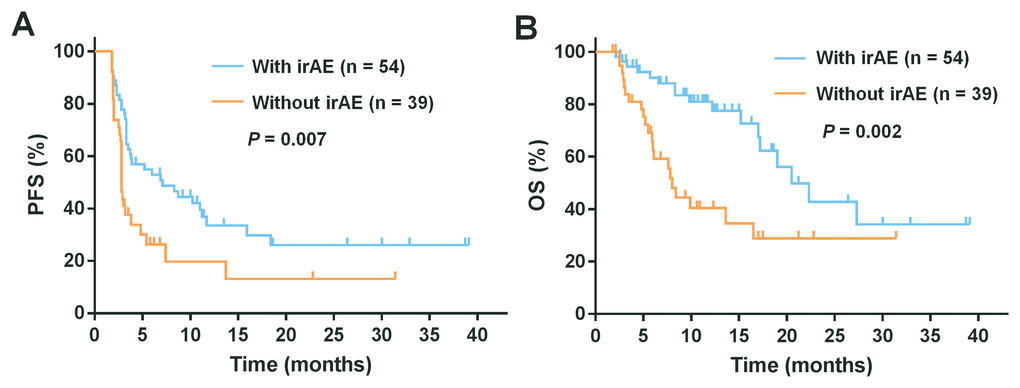
Figure 1. Kaplan-Meier analysis of survival among patients who experienced an immune-related adverse events (irAEs) or not. Shown are the curves for (A) progression-free survival (PFS) and (B) overall survival (OS) in patients with irAEs or not. A statistically significant OS and PFS difference were noted among those experiencing any irAEs versus those who did not (P < 0.05).
The analysis of the association between clinical outcomes and common irAEs revealed that increased PFS was significantly associated with skin irAEs (median 11.0 months; 95% CI, 6.5-15.5 versus 2.8 months; 95% CI, 2.7-2.9, P < 0.001), endocrine irAEs (median Not reached (NR); 95% CI, NR-NR versus 3.3 months; 95% CI, 2.7-3.9, P = 0.006), and fatigue irAEs (median 18.4 months; 95% CI, 4.1-32.7 versus 3.3 months; 95% CI, 2.8-3.8, P = 0.015, respectively). Similarly, increased OS was also significantly associated with skin irAEs (median 22.3 months; 95% CI, NR-NR versus 8.4 months; 95% CI, 5.6-11.2, P < 0.001), endocrine irAEs (median 27.3 months; 95% CI, NR-NR versus 16.5 months; 95% CI, 12.7-20.3, P = 0.047) and fatigue (median NR; 95% CI, NR-NR versus 16.5 months; 95% CI, 13.3-21.7, P = 0.01) (Figure 2A, 2B, and 2E). In contrast, no differences in PFS and OS were observed between patients with and without hepatobiliary and gastrointestinal irAEs (Figure 2C and 2D). Additionally, we also assessed the association between the numbers and grades of irAEs and the prognosis in patients. Patients with three or more irAEs when compared with those with none showed a longer PFS (median 18.4 months; 95% CI, NR-NR versus 2.8 months; 95% CI, 2.7-2.9, P < 0.001) and OS (median NR; 95% CI, NR-NR versus 8.0 months; 95% CI, 6.7-9.3, P < 0.001). Similarly, patients with three or more irAEs when compared with those with one to two irAEs also showed longer PFS (median 18.4 months; 95% CI, NR-NR versus 3.3 months; 95% CI, 2.6-4.0, P < 0.001) and OS (NR; 95% CI, NR-NR versus 19.0 months; 95% CI, 10.1-27.9, P = 0.026) (Figure 3A). However, there were no statistically significant differences in the PFS and OS in patients with one to two irAEs when compared with those with no irAEs (Figure 3A). In addition, patients with grade 1 to 2 irAEs when compared with those with no irAEs showed longer PFS (median 8.7 months; 95% CI, 3.1-14.3 versus 2.8 months; 95% CI, 2.7-2.9, P = 0.005) and OS (22.3 months; 95% CI, 10.6-34.0 versus 8.0 months; 95% CI, 6.7-9.3 P = 0.001) (Figure 3B). There were no significant differences in the PFS and OS of patients with grade 3 to 4 irAEs when compared with those with grade 1 to 2 or no irAEs (Figure 3B). Moreover, the effects of irAEs on the prognosis of patients treated with PD-1 inhibitors were further evaluated by the Cox proportional hazards regression analyses. Multivariable analysis revealed that any irAE and the number of irAEs were significantly associated with increased PFS. Increased OS was also significantly associated with any irAE and the number of irAEs (Table 4).
Table 4. Cox proportional hazard regression analysis of the effect of immune-related adverse events development on progression-free survival and overall survival.
| Survival | Univariate analysis | Multivariate analysisb | |||||||||||||||||||||||||||||||||||||||||||||||||||||||||||||||||||||||||||||||||||||||||||||||||
| HR | 95% CI | P | HR | 95% CI | P | ||||||||||||||||||||||||||||||||||||||||||||||||||||||||||||||||||||||||||||||||||||||||||||||
| PFS | |||||||||||||||||||||||||||||||||||||||||||||||||||||||||||||||||||||||||||||||||||||||||||||||||||
| Any irAEs | 0.509 | 0.305-0.849 | 0.01a | 0.521 | 0.309-0.877 | 0.014a | |||||||||||||||||||||||||||||||||||||||||||||||||||||||||||||||||||||||||||||||||||||||||||||
| Skin irAEs | 0.298 | 0.174-0.510 | <0.001a | 0.297 | 0.172-0.513 | <0.001a | |||||||||||||||||||||||||||||||||||||||||||||||||||||||||||||||||||||||||||||||||||||||||||||
| Endocrine irAEs | 0.232 | 0.073-0.742 | 0.014a | 0.269 | 0.083-0.874 | 0.029a | |||||||||||||||||||||||||||||||||||||||||||||||||||||||||||||||||||||||||||||||||||||||||||||
| Hepatobiliary irAEs | 0.688 | 0.375-1.263 | 0.228 | ||||||||||||||||||||||||||||||||||||||||||||||||||||||||||||||||||||||||||||||||||||||||||||||||
| Gastrointestinal irAEs | 0.939 | 0.347-2.544 | 0.902 | ||||||||||||||||||||||||||||||||||||||||||||||||||||||||||||||||||||||||||||||||||||||||||||||||
| Fatigue irAEs | 0.390 | 0.176-0.899 | 0.021a | 0.410 | 0.181-0.927 | 0.032a | |||||||||||||||||||||||||||||||||||||||||||||||||||||||||||||||||||||||||||||||||||||||||||||
| IrAEs number | 0.532 | 0.384-0.736 | <0.001a | 0.547 | 0.389-0.769 | 0.001a | |||||||||||||||||||||||||||||||||||||||||||||||||||||||||||||||||||||||||||||||||||||||||||||
| irAEs grade | 0.678 | 0.438-1.049 | 0.081 | ||||||||||||||||||||||||||||||||||||||||||||||||||||||||||||||||||||||||||||||||||||||||||||||||
| OS | |||||||||||||||||||||||||||||||||||||||||||||||||||||||||||||||||||||||||||||||||||||||||||||||||||
| Any irAEs | 0.366 | 0.191-0.699 | 0.002a | 0.462 | 0.235-0.909 | 0.025a | |||||||||||||||||||||||||||||||||||||||||||||||||||||||||||||||||||||||||||||||||||||||||||||
| Skin irAEs | 0.257 | 0.124-0.535 | <0.001a | 0.288 | 0.132-0.628 | 0.002a | |||||||||||||||||||||||||||||||||||||||||||||||||||||||||||||||||||||||||||||||||||||||||||||
| Endocrine irAEs | 0.258 | 0.061-1.087 | 0.065 | ||||||||||||||||||||||||||||||||||||||||||||||||||||||||||||||||||||||||||||||||||||||||||||||||
| Hepatobiliary irAEs | 0.497 | 0.195-1.264 | 0.142 | ||||||||||||||||||||||||||||||||||||||||||||||||||||||||||||||||||||||||||||||||||||||||||||||||
| Gastrointestinal irAEs | 0.806 | 0.217-2.991 | 0.747 | ||||||||||||||||||||||||||||||||||||||||||||||||||||||||||||||||||||||||||||||||||||||||||||||||
| Fatigue irAEs | 0.186 | 0.044-0.779 | 0.021a | 0.252 | 0.085-1.092 | 0.065 | |||||||||||||||||||||||||||||||||||||||||||||||||||||||||||||||||||||||||||||||||||||||||||||
| IrAEs number | 0.450 | 0.288-0.705 | <0.001a | 0.529 | 0.326-0.857 | 0.01a | |||||||||||||||||||||||||||||||||||||||||||||||||||||||||||||||||||||||||||||||||||||||||||||
| IrAEs grade | 0.514 | 0.293-0.903 | 0.021a | 0.641 | 0.357-1.151 | 0.136 | |||||||||||||||||||||||||||||||||||||||||||||||||||||||||||||||||||||||||||||||||||||||||||||
| Abbreviations: irAEs, immune-related adverse events; PFS, progression-free survival; OS, overall survival; HR, hazard ratio, CI, confidence interval | |||||||||||||||||||||||||||||||||||||||||||||||||||||||||||||||||||||||||||||||||||||||||||||||||||
| aP value < 0.05 | |||||||||||||||||||||||||||||||||||||||||||||||||||||||||||||||||||||||||||||||||||||||||||||||||||
| b Covariables included metastasis stage (yes versus no), LDH levels (≤UNL versus>UNL), and liver metastases (yes versus no). | |||||||||||||||||||||||||||||||||||||||||||||||||||||||||||||||||||||||||||||||||||||||||||||||||||
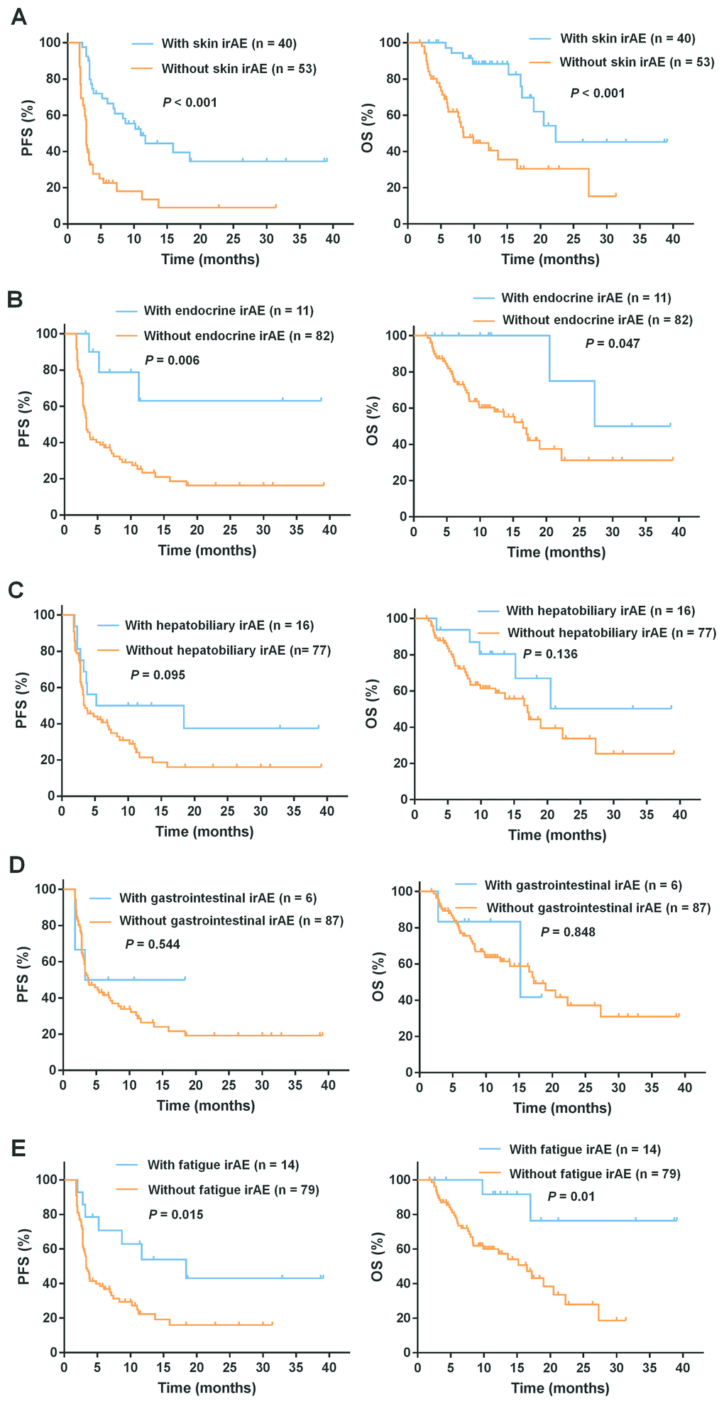
Figure 2. Subgroup analysis to evaluate the association between common irAEs and survival. Shown are the PFS and OS curves for patients with or without (A) skin irAEs, (B) endocrine irAEs, (C) hepatobiliary irAEs, (D) gastrointestinal irAEs, and (E) fatigue. The PFS and OS were significantly associated with skin, endocrine, and fatigue irAEs (P < 0.05).
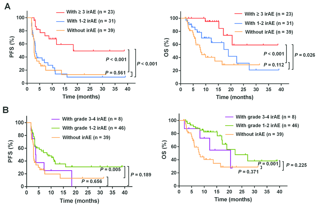
Figure 3. Subgroup analysis to evaluate the association between the number or grade of the irAEs and the prognosis. Shown are the PFS and OS curves for patients with irAEs of different (A) numbers and (B) grades. Patients with three or more irAEs showed longer PFS and OS when compared with those with one to two irAEs or none (P < 0.05). Patients with grade 1 to 2 irAEs showed longer PFS and OS when compared with those with no irAEs (P < 0.01).
Discussion
In this study, we retrospectively analyzed the irAE profiles of PD-1 inhibitors including pembrolizumab and nivolumab. Using a relatively large sample size, we demonstrate that irAEs occurring before disease progression are associated with clinical outcomes in Chinese patients with advanced melanoma. Among the 93 patients, we found that 58.1% of them experienced irAEs with a potential immunological etiology. Most of the irAEs were typically mild to moderate in intensity, and only 8.6% of the patients experienced severe irAEs (grade 3 to 4). Consistent with previous reports [14, 28], the most commonly observed irAEs were rash, pruritus, vitiligo, thyroiditis, hepatitis, fatigue, and pyrexia. However, the incidence rates of certain types of irAEs were slightly different from those previously reported. In our study, after the commencement of treatment with PD-1 inhibitors, irAEs such as pruritus, rash, vitiligo, and hepatitis occurred in 30.1%, 24.7%, 16.1% and 8.6% of patients, respectively. These proportions are marginally higher than those reported in previous clinical trials (14.1-21%, 12-15%, 6-10.7%, and 0-1.1%, respectively) [4, 34–36]. However, the incidence rates of diarrhea and fatigue (5.4% and 15.1 %, respectively) in our study were lower than those previously reported (8-16.9% and 19.9-25%, respectively) [4, 34–36]. One reason for this discrepancy is the different ethnicity, and thereby leads to differences in the baseline characteristics of patients in our study compared with those in other studies. Compared with our study, there were relatively fewer Chinese patients enrolled in the KEYNOTE-001, KEYNOTE-002, KEYNOTE-006, CheckMate-037, and CheckMate-066 trials, which could account for the differences in the irAE profiles.
It has been proposed that the development of irAEs is associated with response to ICIs. This phenomenon was first described in melanoma patients who received ipilimumab therapy [24]. More recently, a retrospective analysis of 576 patients with advanced melanoma received nivolumab monotherapy in United States found a significantly higher ORR in patients who experienced irAEs of any grade than those who did not, with greater benefit in patients with three or more or one to two treatment-related AEs than those with none [27]. Consistent with these findings, our study also demonstrated that the ORR and DCR were significantly better in patients who experienced irAEs of any grade than those who did not. However, we found stronger associations between clinical outcomes and those with three or more irAEs than those with less than three irAEs. We also found that while the ORR and DCR were significantly higher in patients with grade 1 to 2 irAEs than those with no irAEs, but no significantly better outcomes were observed in patients with grade 3 to 4 irAEs than those with no irAEs. Furthermore, we found that patients with grade 3 to 4 irAEs showed poorer clinical outcomes when compared with those with grade 1 to 2 irAEs. A recent retrospective study also found that while ORR was significantly associated with low-grade irAEs in non-melanoma patients treated with PD-1 inhibitors, no significant relationship was found with high-grade irAEs [37]. The poorer clinical response in patients who developed grade 3 to 4 irAEs is potentially due to the termination of immunotherapy when faced with irAEs and the use of steroids.
Prior studies have demonstrated an association between the development of irAEs and clinical survival in tumor patients treated with PD-1 inhibitors. Analysis of 148 melanoma patients treated with nivolumab in the United States, the OS was greater in patients with irAEs especially three or more than those who had no irAEs [25]. A previous prospective study also reported that early irAEs are associated with a better PFS in NSCLC patients receiving nivolumab treatment [18]. Another retrospective study analyzed NSCLC patients treated with pembrolizumab from a single center in the KEYNOTE-001 trial and found that the patients who experienced irAEs had increased PFS and OS, compared to those who did not [20]. Consistent with these previous studies, we also found that the development of irAEs was significantly associated with increased PFS and OS, particularly in patients with three or more irAEs. In addition, we found that prolonged survival of advanced melanoma patients treated with PD-1 inhibitors was associated with skin and endocrine irAEs, as well as fatigue, but not with hepatobiliary and gastrointestinal irAEs. Our findings are consistent with some other studies that have showed an association between skin irAEs and prolonged survival in patients with NSCLC and melanoma treated with nivolumab [25, 28]. Moreover, a previous study on malignant melanoma revealed clear survival benefits in patients who received immunotherapy and developed vitiligo-like depigmentation [26]. In contrast to our results, it has been reported that hypothyroidism or hyperthyroidism was not associated with prolonged survival in malignant melanoma [25]. However, thyroid dysfunction was associated with improved OS of NSCLC patients treated with pembrolizumab [12].
The mechanism underlying the association of irAEs with the efficacy and clinical outcomes of treatment with PD-1 inhibitors remains unclear. However, previous studies have demonstrated that melanocytes and melanomas shared common antigens (e.g., MART-1, gp100, and tyrosinase related proteins 1 and 2) and lymphocytes directed against the tumor could cross-react with normal melanocytes and cause irAEs in the skin [11, 13, 28, 38]. In view of these findings, antigen sharing was considered the most likely cause of this association. Therefore, we speculate that irAEs can predict a strong activation of the immune system following the inhibition of PD-1, which can explain the better prognosis in melanoma patients, give the sensitivity of these tumors to immunotherapy.
In conclusion, this retrospective study demonstrated that irAEs were associated with the efficacy of PD-1 inhibitors in Chinese patients with advanced melanoma. Moreover, we showed that patients with at least three irAEs of grade 1 or 2, which include dermal, endocrine and fatigue were most likely to benefit from the inhibition of PD-1. However, our findings should be validated prospectively in subsequent analyses in larger cohorts of patients with advanced melanoma.
Materials and Methods
Patient population
We performed a retrospective study of Chinese patients with advanced melanoma who received PD-1 inhibitor monotherapy at the Sun Yat-sen University Cancer Center between August 2014 and March 2018. Patients with histologically confirmed advanced melanoma derived from skin and non-skin sections were included in this study. The included patients were treated with intravenous pembrolizumab (2 mg/kg every 3 weeks) or nivolumab (3mg/kg every 2 weeks). After excluding 11 patients who received only one dose of the drug, and 2 patients who died before the first imaging evaluation, a total of 93 patients were finally enrolled in our study. The electronic medical records were reviewed to obtain information including patient demographic, ECOG status, primary site, metastasis stage, BRAF V600E status, prior therapy, number of anti-PD-1 drug doses received, any irAEs, use of corticosteroids, findings of the imaging evaluation, date of progression and start of new treatment or death. The follow-up was ended on June 1, 2018. The Institutional Review Board of the Sun Yat-sen University Cancer Center approved the study, and all the patients provided written informed consent.
Assessments
IrAEs are defined as AEs with a potential immunologic cause that require frequent monitoring and intervention with immunosuppressive and/or endocrine therapy [5, 39–44]. All irAEs were graded according to the National Cancer Institute Common Terminology Criteria for Adverse Events (version 4.0). Radiological evaluations (CT or MRI) were performed at baseline and subsequently at every 8 to 12 weeks to assess tumor responses. Tumor assessments were made based on the Response Evaluation Criteria in Solid Tumors (RECIST version 1.1), and included complete remission (CR), partial remission (PR), stable disease (SD), and progressive disease (PD). The ORR (CR + PR) and DCR (CR + PR + SD) were also calculated. PFS was defined as the interval between the start of the treatment and disease progression or death due to any cause. OS was defined as the interval between the start of the treatment and death due to any cause. Patients who did not progress or were still alive at the last follow-up date were censored.
Statistical analysis
Clinical and demographic characteristics of the patient with and without irAEs were compared using the Fisher exact test for categorical variables. ORRs and DCRs with 95% CIs were estimated using the Clopper-Pearson method. The Fisher exact tests were used to determine the associations between the number/grade of irAEs and the tumor response rates including ORRs and DCRs. The differences in the PFS and OS curves (estimated by the Kaplan-Meier method) based on the absence or presence of any irAEs which were observed before disease progression was evaluated using the log-rank test. We used a Cox proportional hazards model to calculate HRs and 95% CIs. A multivariable analysis was performed with adjustment for metastasis stage, LDH levels, and liver metastases. A two-tailed p value < 0.05 was considered to be statistically significant. All the statistical analyses were performed using the IBM SPSS 19.0 software.
Abbreviations
PD-1: programmed cell death 1; irAEs: immune-related adverse events; ORR: overall response rate; DCR: disease control rate; PFS: progression-free survival; OS: overall survival; ICIs: immune checkpoint inhibitors; NSCLC: non–small cell lung cancer; CSD: chronic sun-derived; ORR: the objective response rate; DCR: disease control rate; PFS: progression-free survival; OS: overall survival; NR: Not reached; CR: complete remission; PR: partial remission; SD: stable disease; PD: progressive disease.
Acknowledgments
The authors would like to thank and acknowledge all patients for taking part in this study and their families.
Conflicts of Interest
The authors declare that they have no competing interests.
Funding
This work was supported by grants from the National Natural Science Foundation of China (No. 81402560; 81772910; 81572493), the Guangdong Province Science and Technology Plan Project (No. 2017A020215029) and Guangdong Esophageal Cancer Institute Science and Technology Program (No. Q201802).
References
- 1. Ferlay J, Soerjomataram I, Dikshit R, Eser S, Mathers C, Rebelo M, Parkin DM, Forman D, Bray F. Cancer incidence and mortality worldwide: sources, methods and major patterns in GLOBOCAN 2012. Int J Cancer. 2015; 136:E359–86. https://doi.org/10.1002/ijc.29210 [PubMed]
- 2. Wen X, Wang Y, Ding Y, Li D, Li J, Guo Y, Peng R, Zhao J, Zhang X, Zhang XS. Safety of immune checkpoint inhibitors in chinese patients with melanoma. Melanoma Res. 2016; 26:284–89. https://doi.org/10.1097/CMR.0000000000000256 [PubMed]
- 3. McArthur GA, Ribas A. Targeting oncogenic drivers and the immune system in melanoma. J Clin Oncol. 2013; 31:499–506. https://doi.org/10.1200/JCO.2012.45.5568 [PubMed]
- 4. Ribas A, Puzanov I, Dummer R, Schadendorf D, Hamid O, Robert C, Hodi FS, Schachter J, Pavlick AC, Lewis KD, Cranmer LD, Blank CU, O’Day SJ, et al. Pembrolizumab versus investigator-choice chemotherapy for ipilimumab-refractory melanoma (KEYNOTE-002): a randomised, controlled, phase 2 trial. Lancet Oncol. 2015; 16:908–18. https://doi.org/10.1016/S1470-2045(15)00083-2 [PubMed]
- 5. Chen TW, Razak AR, Bedard PL, Siu LL, Hansen AR. A systematic review of immune-related adverse event reporting in clinical trials of immune checkpoint inhibitors. Ann Oncol. 2015; 26:1824–29. https://doi.org/10.1093/annonc/mdv182 [PubMed]
- 6. Larkin J, Chiarion-Sileni V, Gonzalez R, Grob JJ, Cowey CL, Lao CD, Schadendorf D, Dummer R, Smylie M, Rutkowski P, Ferrucci PF, Hill A, Wagstaff J, et al. Combined Nivolumab and Ipilimumab or Monotherapy in Untreated Melanoma. N Engl J Med. 2015; 373:23–34. https://doi.org/10.1056/NEJMoa1504030 [PubMed]
- 7. Robert C, Ribas A, Wolchok JD, Hodi FS, Hamid O, Kefford R, Weber JS, Joshua AM, Hwu WJ, Gangadhar TC, Patnaik A, Dronca R, Zarour H, et al. Anti-programmed-death-receptor-1 treatment with pembrolizumab in ipilimumab-refractory advanced melanoma: a randomised dose-comparison cohort of a phase 1 trial. Lancet. 2014; 384:1109–17. https://doi.org/10.1016/S0140-6736(14)60958-2 [PubMed]
- 8. Weide B, Martens A, Hassel JC, Berking C, Postow MA, Bisschop K, Simeone E, Mangana J, Schilling B, Di Giacomo AM, Brenner N, Kähler K, Heinzerling L, et al. Baseline biomarkers for outcome of melanoma patients treated with pembrolizumab. Clin Cancer Res. 2016; 22:5487–96. https://doi.org/10.1158/1078-0432.CCR-16-0127 [PubMed]
- 9. Hassel JC, Heinzerling L, Aberle J, Bähr O, Eigentler TK, Grimm MO, Grünwald V, Leipe J, Reinmuth N, Tietze JK, Trojan J, Zimmer L, Gutzmer R. Combined immune checkpoint blockade (anti-PD-1/anti-CTLA-4): evaluation and management of adverse drug reactions. Cancer Treat Rev. 2017; 57:36–49. https://doi.org/10.1016/j.ctrv.2017.05.003 [PubMed]
- 10. Hamid O, Robert C, Daud A, Hodi FS, Hwu WJ, Kefford R, Wolchok JD, Hersey P, Joseph RW, Weber JS, Dronca R, Gangadhar TC, Patnaik A, et al. Safety and tumor responses with lambrolizumab (anti-PD-1) in melanoma. N Engl J Med. 2013; 369:134–44. https://doi.org/10.1056/NEJMoa1305133 [PubMed]
- 11. Hua C, Boussemart L, Mateus C, Routier E, Boutros C, Cazenave H, Viollet R, Thomas M, Roy S, Benannoune N, Tomasic G, Soria JC, Champiat S, et al. Association of vitiligo with tumor response in patients with metastatic melanoma treated with pembrolizumab. JAMA Dermatol. 2016; 152:45–51. https://doi.org/10.1001/jamadermatol.2015.2707 [PubMed]
- 12. Osorio JC, Ni A, Chaft JE, Pollina R, Kasler MK, Stephens D, Rodriguez C, Cambridge L, Rizvi H, Wolchok JD, Merghoub T, Rudin CM, Fish S, Hellmann MD. Antibody-mediated thyroid dysfunction during t-cell checkpoint blockade in patients with non-small-cell lung cancer. Ann Oncol. 2017; 28:583–89. https://doi.org/10.1093/annonc/mdw640 [PubMed]
- 13. Sanlorenzo M, Vujic I, Daud A, Algazi A, Gubens M, Luna SA, Lin K, Quaglino P, Rappersberger K, Ortiz-Urda S. Pembrolizumab cutaneous adverse events and their association with disease progression. JAMA Dermatol. 2015; 151:1206–12. https://doi.org/10.1001/jamadermatol.2015.1916 [PubMed]
- 14. Topalian SL, Sznol M, McDermott DF, Kluger HM, Carvajal RD, Sharfman WH, Brahmer JR, Lawrence DP, Atkins MB, Powderly JD, Leming PD, Lipson EJ, Puzanov I, et al. Survival, durable tumor remission, and long-term safety in patients with advanced melanoma receiving nivolumab. J Clin Oncol. 2014; 32:1020–30. https://doi.org/10.1200/JCO.2013.53.0105 [PubMed]
- 15. Topalian SL, Hodi FS, Brahmer JR, Gettinger SN, Smith DC, McDermott DF, Powderly JD, Carvajal RD, Sosman JA, Atkins MB, Leming PD, Spigel DR, Antonia SJ, et al. Safety, activity, and immune correlates of anti-PD-1 antibody in cancer. N Engl J Med. 2012; 366:2443–54. https://doi.org/10.1056/NEJMoa1200690 [PubMed]
- 16. Weber JS, Kudchadkar RR, Yu B, Gallenstein D, Horak CE, Inzunza HD, Zhao X, Martinez AJ, Wang W, Gibney G, Kroeger J, Eysmans C, Sarnaik AA, Chen YA. Safety, efficacy, and biomarkers of nivolumab with vaccine in ipilimumab-refractory or -naive melanoma. J Clin Oncol. 2013; 31:4311–18. https://doi.org/10.1200/JCO.2013.51.4802 [PubMed]
- 17. Wolchok JD, Kluger H, Callahan MK, Postow MA, Rizvi NA, Lesokhin AM, Segal NH, Ariyan CE, Gordon RA, Reed K, Burke MM, Caldwell A, Kronenberg SA, et al. Nivolumab plus ipilimumab in advanced melanoma. N Engl J Med. 2013; 369:122–33. https://doi.org/10.1056/NEJMoa1302369 [PubMed]
- 18. Teraoka S, Fujimoto D, Morimoto T, Kawachi H, Ito M, Sato Y, Nagata K, Nakagawa A, Otsuka K, Uehara K, Imai Y, Ishida K, Fukuoka J, Tomii K. Early immune-related adverse events and association with outcome in advanced non-small cell lung cancer patients treated with nivolumab: a prospective cohort study. J Thorac Oncol. 2017; 12:1798–805. https://doi.org/10.1016/j.jtho.2017.08.022 [PubMed]
- 19. Champiat S, Lambotte O, Barreau E, Belkhir R, Berdelou A, Carbonnel F, Cauquil C, Chanson P, Collins M, Durrbach A, Ederhy S, Feuillet S, François H, et al. Management of immune checkpoint blockade dysimmune toxicities: a collaborative position paper. Ann Oncol. 2016; 27:559–74. https://doi.org/10.1093/annonc/mdv623 [PubMed]
- 20. Lisberg A, Tucker DA, Goldman JW, Wolf B, Carroll J, Hardy A, Morris K, Linares P, Adame C, Spiegel ML, Wells C, McKenzie J, Ledezma B, et al. Treatment-related adverse events predict improved clinical outcome in NSCLC patients on KEYNOTE-001 at a single center. Cancer Immunol Res. 2018; 6:288–94. https://doi.org/10.1158/2326-6066.CIR-17-0063 [PubMed]
- 21. Naidoo J, Page DB, Li BT, Connell LC, Schindler K, Lacouture ME, Postow MA, Wolchok JD. Toxicities of the anti-PD-1 and anti-PD-L1 immune checkpoint antibodies. Ann Oncol. 2015; 26:2375–91. https://doi.org/10.1093/annonc/mdv383 [PubMed]
- 22. Spain L, Diem S, Larkin J. Management of toxicities of immune checkpoint inhibitors. Cancer Treat Rev. 2016; 44:51–60. https://doi.org/10.1016/j.ctrv.2016.02.001 [PubMed]
- 23. Voskens CJ, Goldinger SM, Loquai C, Robert C, Kaehler KC, Berking C, Bergmann T, Bockmeyer CL, Eigentler T, Fluck M, Garbe C, Gutzmer R, Grabbe S, et al. The price of tumor control: an analysis of rare side effects of anti-CTLA-4 therapy in metastatic melanoma from the ipilimumab network. PLoS One. 2013; 8:e53745. https://doi.org/10.1371/journal.pone.0053745 [PubMed]
- 24. Downey SG, Klapper JA, Smith FO, Yang JC, Sherry RM, Royal RE, Kammula US, Hughes MS, Allen TE, Levy CL, Yellin M, Nichol G, White DE, et al. Prognostic factors related to clinical response in patients with metastatic melanoma treated by CTL-associated antigen-4 blockade. Clin Cancer Res. 2007; 13:6681–88. https://doi.org/10.1158/1078-0432.CCR-07-0187 [PubMed]
- 25. Freeman-Keller M, Kim Y, Cronin H, Richards A, Gibney G, Weber JS. Nivolumab in resected and unresectable metastatic melanoma: characteristics of immune-related adverse events and association with outcomes. Clin Cancer Res. 2016; 22:886–94. https://doi.org/10.1158/1078-0432.CCR-15-1136 [PubMed]
- 26. Teulings HE, Limpens J, Jansen SN, Zwinderman AH, Reitsma JB, Spuls PI, Luiten RM. Vitiligo-like depigmentation in patients with stage III-IV melanoma receiving immunotherapy and its association with survival: a systematic review and meta-analysis. J Clin Oncol. 2015; 33:773–81. https://doi.org/10.1200/JCO.2014.57.4756 [PubMed]
- 27. Weber JS, Hodi FS, Wolchok JD, Topalian SL, Schadendorf D, Larkin J, Sznol M, Long GV, Li H, Waxman IM, Jiang J, Robert C. Safety profile of nivolumab monotherapy: a pooled analysis of patients with advanced melanoma. J Clin Oncol. 2017; 35:785–92. https://doi.org/10.1200/JCO.2015.66.1389 [PubMed]
- 28. Haratani K, Hayashi H, Chiba Y, Kudo K, Yonesaka K, Kato R, Kaneda H, Hasegawa Y, Tanaka K, Takeda M, Nakagawa K. Association of immune-related adverse events with nivolumab efficacy in non-small-cell lung cancer. JAMA Oncol. 2018; 4:374–78. https://doi.org/10.1001/jamaoncol.2017.2925 [PubMed]
- 29. Hasan Ali O, Diem S, Markert E, Jochum W, Kerl K, French LE, Speiser DE, Früh M, Flatz L. Characterization of nivolumab-associated skin reactions in patients with metastatic non-small cell lung cancer. Oncoimmunology. 2016; 5:e1231292. https://doi.org/10.1080/2162402X.2016.1231292 [PubMed]
- 30. Curtin JA, Fridlyand J, Kageshita T, Patel HN, Busam KJ, Kutzner H, Cho KH, Aiba S, Bröcker EB, LeBoit PE, Pinkel D, Bastian BC. Distinct sets of genetic alterations in melanoma. N Engl J Med. 2005; 353:2135–47. https://doi.org/10.1056/NEJMoa050092 [PubMed]
- 31. Furney SJ, Turajlic S, Stamp G, Nohadani M, Carlisle A, Thomas JM, Hayes A, Strauss D, Gore M, van den Oord J, Larkin J, Marais R. Genome sequencing of mucosal melanomas reveals that they are driven by distinct mechanisms from cutaneous melanoma. J Pathol. 2013; 230:261–69. https://doi.org/10.1002/path.4204 [PubMed]
- 32. Furney SJ, Turajlic S, Stamp G, Thomas JM, Hayes A, Strauss D, Gavrielides M, Xing W, Gore M, Larkin J, Marais R. The mutational burden of acral melanoma revealed by whole-genome sequencing and comparative analysis. Pigment Cell Melanoma Res. 2014; 27:835–38. https://doi.org/10.1111/pcmr.12279 [PubMed]
- 33. Wen X, Ding Y, Li J, Zhao J, Peng R, Li D, Zhu B, Wang Y, Zhang X, Zhang X. The experience of immune checkpoint inhibitors in chinese patients with metastatic melanoma: a retrospective case series. Cancer Immunol Immunother. 2017; 66:1153–62. https://doi.org/10.1007/s00262-017-1989-8 [PubMed]
- 34. Robert C, Long GV, Brady B, Dutriaux C, Maio M, Mortier L, Hassel JC, Rutkowski P, McNeil C, Kalinka-Warzocha E, Savage KJ, Hernberg MM, Lebbé C, et al. Nivolumab in previously untreated melanoma without BRAF mutation. N Engl J Med. 2015; 372:320–30. https://doi.org/10.1056/NEJMoa1412082 [PubMed]
- 35. Robert C, Schachter J, Long GV, Arance A, Grob JJ, Mortier L, Daud A, Carlino MS, McNeil C, Lotem M, Larkin J, Lorigan P, Neyns B, et al, and KEYNOTE-006 investigators. Pembrolizumab versus ipilimumab in advanced melanoma. N Engl J Med. 2015; 372:2521–32. https://doi.org/10.1056/NEJMoa1503093 [PubMed]
- 36. Weber JS, D'Angelo SP, Minor D, Hodi FS, Gutzmer R, Neyns B, Hoeller C, Khushalani NI, Miller WH
Jr , Lao CD, Linette GP, Thomas L, Lorigan P, et al. Nivolumab versus chemotherapy in patients with advanced melanoma who progressed after anti-CTLA-4 treatment (CheckMate 037): a randomised, controlled, open-label, phase 3 trial. Lancet Oncol. 2015; 16:375–84. https://doi.org/10.1016/S1470-2045(15)70076-8 [PubMed] - 37. Judd J, Zibelman M, Handorf E, O’Neill J, Ramamurthy C, Bentota S, Doyle J, Uzzo RG, Bauman J, Borghaei H, Plimack ER, Mehra R, Geynisman DM. Immune-related adverse events as a biomarker in non-melanoma patients treated with programmed cell death 1 inhibitors. Oncologist. 2017; 22:1232–37. https://doi.org/10.1634/theoncologist.2017-0133 [PubMed]
- 38. Houghton AN, Eisinger M, Albino AP, Cairncross JG, Old LJ. Surface antigens of melanocytes and melanomas. Markers of melanocyte differentiation and melanoma subsets. J Exp Med. 1982; 156:1755–66. https://doi.org/10.1084/jem.156.6.1755 [PubMed]
- 39. Borghaei H, Paz-Ares L, Horn L, Spigel DR, Steins M, Ready NE, Chow LQ, Vokes EE, Felip E, Holgado E, Barlesi F, Kohlhäufl M, Arrieta O, et al. Nivolumab versus docetaxel in advanced nonsquamous non-small-cell lung cancer. N Engl J Med. 2015; 373:1627–39. https://doi.org/10.1056/NEJMoa1507643 [PubMed]
- 40. Boutros C, Tarhini A, Routier E, Lambotte O, Ladurie FL, Carbonnel F, Izzeddine H, Marabelle A, Champiat S, Berdelou A, Lanoy E, Texier M, Libenciuc C, et al. Safety profiles of anti-CTLA-4 and anti-PD-1 antibodies alone and in combination. Nat Rev Clin Oncol. 2016; 13:473–86. https://doi.org/10.1038/nrclinonc.2016.58 [PubMed]
- 41. Brahmer J, Reckamp KL, Baas P, Crinò L, Eberhardt WE, Poddubskaya E, Antonia S, Pluzanski A, Vokes EE, Holgado E, Waterhouse D, Ready N, Gainor J, et al. Nivolumab versus docetaxel in advanced squamous-cell non-small-cell lung cancer. N Engl J Med. 2015; 373:123–35. https://doi.org/10.1056/NEJMoa1504627 [PubMed]
- 42. Herbst RS, Baas P, Kim DW, Felip E, Pérez-Gracia JL, Han JY, Molina J, Kim JH, Arvis CD, Ahn MJ, Majem M, Fidler MJ, de Castro G
Jr , et al. Pembrolizumab versus docetaxel for previously treated, PD-L1-positive, advanced non-small-cell lung cancer (KEYNOTE-010): a randomised controlled trial. Lancet. 2016; 387:1540–50. https://doi.org/10.1016/S0140-6736(15)01281-7 [PubMed] - 43. Reck M, Rodríguez-Abreu D, Robinson AG, Hui R, Csőszi T, Fülöp A, Gottfried M, Peled N, Tafreshi A, Cuffe S, O'Brien M, Rao S, Hotta K, et al, and KEYNOTE-024 Investigators. Pembrolizumab versus chemotherapy for PD-L1-positive non-small-cell lung cancer. N Engl J Med. 2016; 375:1823–33. https://doi.org/10.1056/NEJMoa1606774 [PubMed]
- 44. Yamazaki N, Kiyohara Y, Uhara H, Fukushima S, Uchi H, Shibagaki N, Tsutsumida A, Yoshikawa S, Okuyama R, Ito Y, Tokudome T. Phase II study of ipilimumab monotherapy in Japanese patients with advanced melanoma. Cancer Chemother Pharmacol. 2015; 76:997–1004. https://doi.org/10.1007/s00280-015-2873-x [PubMed]