


Introduction
Colon cancer is a common cause of morbidity and mortality worldwide [1]. There were an estimated 101420 new cases of colon cancer in the US in 2019, and an estimated 44180 patients died from colorectal cancer in the same year [2]. For resectable nonmetastatic colon cancer, the preferred treatment method is colectomy plus regional lymph node dissection [3]. Unfortunately, approximately 50%-60% of patients diagnosed with colon cancer develop metastases, and of these, 80% to 90% are nonresectable liver metastases with poor prognosis [4, 5]. In recent years, colon cancer patients have benefited significantly from 5-Fu- and oxaliplatin-based chemotherapy regimens, and newly developed targeted drugs also prolong the survival time of patients with advanced colon cancer [6, 7]. However, recurrence and metastasis remain major problems in colon cancer and are often the ultimate causes of death.
Currently, colon cancer has shifted from the inherent treatment mode of "surgery mainly, chemoradiotherapy supplemented" to the treatment concept of precision and individual, and immunotherapy is receiving increasing attention [8]. In 2015, Le et al. found that metastatic colorectal cancer (mCRC) with either different mismatch repair (dMMR) or highly microsatellite instability (MSI-H) molecular phenotypes can significantly benefit from the immune checkpoint inhibitor (ICPI) programmed death ligand 1 (PD-L1) monoclonal antibody pembrolizumab [9]. Although subsequent studies have further expanded the immunotherapy indications for MSI-H/dMMR colorectal cancer from the original posttreatment of metastatic disease to first-line treatment and neoadjuvant therapy for early disease [10, 11], the effective population of immunotherapy is still limited to this specific group. Currently, immunohistochemical detection of PD-L1 has been widely used to screen patients with colorectal cancer who can benefit from immunotherapy [12]. However, the tumor tissue microenvironment can interfere with PD-L1 expression, and the relationship between the expression of PD-L1 in colorectal cancer and the efficacy of immunotherapy is not exact [13]. Moreover, the response rate of MSI-H colorectal cancer patients to ICPI is also variable, and tumor responders have more somatic mutations and higher neoantigen load than nonresponders [14], indicating the need for additional predictive biomarkers.
Tumor mutant burden (TMB) is a biomarker reflecting somatic mutation and is expected to pave the way for tumor immunotherapy to enter the era of precision medicine [15]. During the occurrence and development of cancer, a mass of somatic mutations can produce neoantigens, which increased tumor immunogenicity and thereby activated immune recognition system [16]. The high production of neoantigens is associated with enhanced checkpoint blocking responses, and along with the recognition of neoantigens, the activity of T cells against the tumor was increased by immune system thereby enhancing the efficacy of ICPI [17–19]. TMB is an emerging independent biomarker that can be used to stratify the possible response of patients to ICPC [20]. A previous study found that among the patients with high TMB lung cancer, the response rate for ICPI was higher than that in the patients with low TMB expression, and the clinical outcome was significantly improved, suggesting that high TMB was positively correlated with the efficacy of immunotherapy [21]. In addition, Zaravinos et al. have reported that colon cancer cells possess a higher mutation load and neoepitope load, which drives the immune system to fight against tumors [22]. However, the changes of specific gene mutations and their relationship with TMB and tumor-infiltrating immune cells in colon cancer remains unclear. Therefore, the aim of our study is to identify mutated genes using TCGA and ICGC colon cancer samples, and to further explore the association of mutated genes with TMB and patient outcome and infiltrating immune cells.
Results
Somatic mutation characteristics in colon cancer
We first downloaded the mutation data of 398 American colon cancer samples from TCGA, and the cumulative mutations frequency in each gene was counted and sorted in decreasing order. The top 30 frequently mutated genes with high mutation frequency and the pattern of somatic mutation for the top 30 genes are illustrated in Figure 1A. The top 5 mutated genes were APC (74%), TP53 (54%), TTN (48%), KRAS (43%), and SYNE1 (29%). Similarly, the top 30 mutated genes were also identified in Chinese patients from ICGC database. As shown in Figure 1B, missense mutation was occurred commonly in Chinese patients, and APC (49%), TP53 (46%), TTN (39%), KRAS (37%), and MUC6 (35%) had the top 5 mutation frequency among Chinese patients.
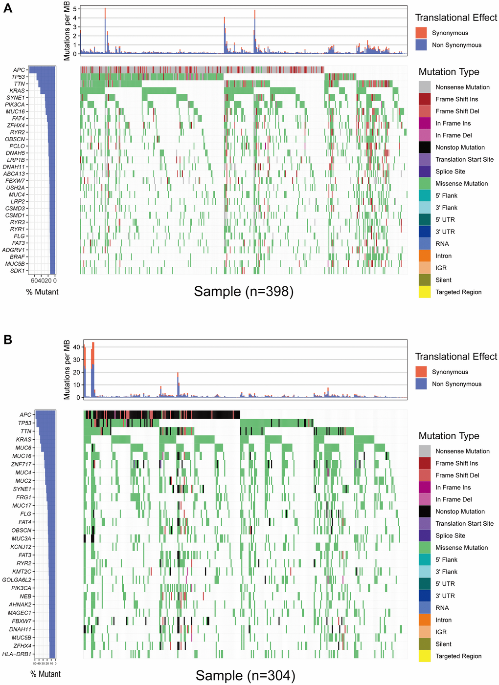
Figure 1. Overview of frequently mutated genes in colon cancer. (A) Waterfall plot shows the frequently mutated genes in colon cancer from TCGA database. The left panel shows mutation frequency, and genes are ordered by their mutation frequencies. The right panel presents different mutation types. (B) Waterfall plot displaying the frequently mutated genes in colon cancer from the ICGC cohort. The left panel shows the genes ordered by their mutation frequencies. The right panel presents different mutation types.
Gene mutations associated with TMB
Next, to obtain the genes that are commonly mutated in both TCGA and ICGC databases. we intersected the genes with the top 30 mutation rates in the two cohorts. As shown in Figure 2A, the intersection genes with high mutations were APC, TP53, TNN, KRAS, MUC16, MUC4, SYNE1, FLG, FAT4, OBSCN, FAT3, RYR2, PIK3CA, FBXW7, DNAH11, MUC5B and ZFHX4. To further investigated whether these 17 commonly mutated genes were associated with TMB, colon cancer patients from TCGA cohort were classified into wild group and mutation group based on the 17 gene mutation status. In addition, TMB expression for each TCGA sample was calculated, and the median value of TMB is 9.95 per Mb (0.05-188.32 per Mb). With combining analysis of the data of gene mutation matrix and TMB expression matrix, we found that TMB value in mutation group of all the other 16 genes except KRAS was significantly changed compared with wild group (Figure 2B).
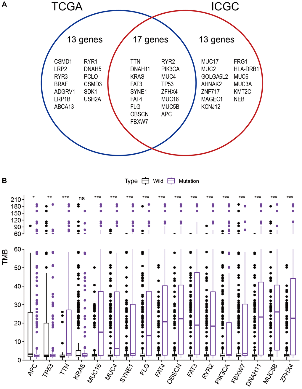
Figure 2. Gene mutations are associated with TMB. (A) Venn diagram shows 17 frequently mutated genes covered by both the TCGA and ICGC cohorts. (B) Sixteen genes with high mutation frequency are associated with a higher TMB. *p < 0.05; **p < 0.01; ***p < 0.001; ns: no significance.
MUC4 mutation associated with prognosis
It is well known that TMB is associated with the relapse-free survival (RFS) in colon cancer [23]. Thus, considering the established association between 16 mutated genes and TMB, we speculate that these genes may be associated with clinical outcomes. For this purpose, patients from TCGA database were assigned to wild group and mutation group according to genes mutation status and Kaplan-Meier analysis was conducted with combining analysis of patient survival data. Our results demonstrated that only MUC4 mutation was associated with a poor prognosis (p = 0.009) (Figure 3). Based on this funding, we aimed to further identify whether MUC4 mutation is the independent prognostic factor for colon cancer using Cox regression analysis. As shown in Figure 4, With correction for common clinical information and TMB score, MUC4 mutation remained significantly associated with overall survival of patients.
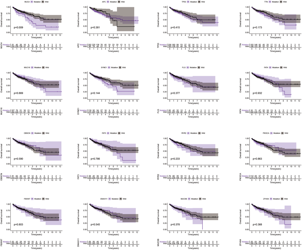
Figure 3. MUC4 mutation is associated with clinical prognosis. Kaplan-Meier survival analysis was used to determine survival curves that reflect the association between gene mutations and prognosis. The p-value is shown each plot.
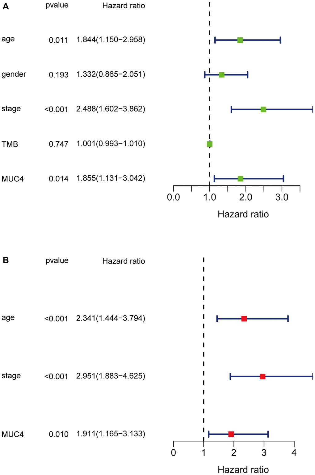
Figure 4. Univariate (A) and multivariate (B) overall survival analysis of colon cancer patients by the Cox proportional hazards model.
Identification of enrichment pathways for patients with MUC4 mutation
We next investigated the enrichment pathway associated with MUC4 mutation. GSEA was performed, and the results showed that pathways were significantly enriched in the MUC4 mutant group, including antigen processing and presentation, cytosolic DNA sensing pathway, prion diseases, graft versus host disease, type I diabetes mellitus, leishmania infection, toll like receptor signaling pathway, natural killer cell mediated cytotoxicity and prostate cancer (Figure 5A). Pathways that were significantly enriched in the MUC4 wild-type group included glycosylphosphatidylinositol GPI anchor biosynthesis, peroxisome, primary bile acid biosynthesis, and riboflavin metabolism (Figure 5B). It is widely recognized that TMB is helpful to screen beneficiaries and predict the effect of immunotherapy. Considering the established association between MUC4 mutation and TMB, thereby we speculate MUC4 mutation may be correlated with immune response. As shown in Figure 5C, we observed that some immune-related pathways, including cytosolic DNA sensing pathway, antigen processing and presentation, natural killer cell mediated cytotoxicity, graft versus host disease and toll like receptor signaling pathway were enriched in MUC4 mutation samples, while no immune response-related pathway was enriched in samples with wild-type MUC4.
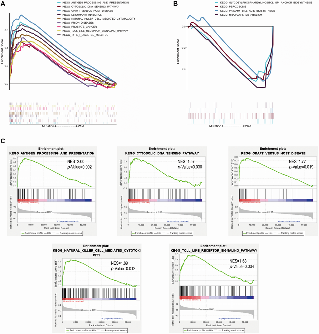
Figure 5. MUC4 mutation is associated with immune-related pathways. Gene set enrichment analysis was performed with the TCGA. (A) Multiple gene enrichment plot shows that a series of gene sets are enriched in the MUC4-mutant group. (B) Multiple gene enrichment plot shows that a series of gene sets are enriched in the wild-type MUC4 group. (C) Gene enrichment plots display that a series of immune-related gene sets are enriched in the MUC4-mutant group. NES, normalized enrichment score. The p-value is shown in each plot.
Tumor-infiltrating immune cells associated with MUC4 mutation in colon cancer
Using CIBERSORT deconvolution algorithm, we first calculated the proportion of 22 immune cells for each sample in tumor tissue. The results revealed that the number of infiltrating immune cells changes greatly in different sample, and T cells and macrophages accounted for a relatively high proportion in the total samples (Figure 6A). Next, these samples were divided into MUC4 wild group and MUC4 mutation group to evaluate the situation of immune cell infiltration in the two groups. Compared to MUC4 wild group, the infiltration proportion of follicular helper T cells and activated memory CD4 T cells were higher in MUC4 mutant group (Figure 6B). Finally, correlation analysis revealed that activated memory CD4 T cells had the strongest positive correlation with CD8 T cells, while they were negatively correlated with resting memory CD4 T cells and Tregs (regulatory T cells) (Figure 6C). Moreover, follicular helper T cells had the strongest positive correlation with CD8 T cells, and had the strongest negative correlation with M0 macrophages (Figure 6C).
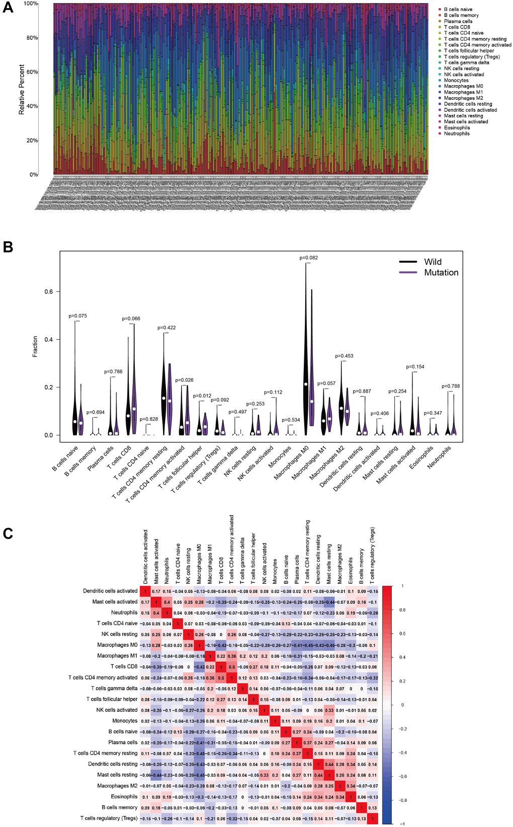
Figure 6. MUC4 mutation is correlated with tumor-infiltrating immune cells. (A) The stacked bar chart shows the distribution of 22 immune cells in each sample. (B) Violin plot displaying the differentially infiltrated immune cells between the MUC4-mutant groups and the wild-type MUC4 group. black represents the wild-type MUC4 group, and purple represents the MUC4-mutant group. The p-value is shown in the figure. (C) Correlation matrix of immune cell proportions. The red color represents a positive correlation, and the blue color represents a negative correlation.
Discussion
In our study, somatic mutation landscapes of colon cancer were characterized in 398 American samples and 304 Chinese samples. Subsequently, MUC4 mutation was identified to be associated with TMB and patient clinical outcomes. Moreover, immune-related signaling pathways were significantly enriched in samples with MUC4 mutation. Furthermore, MUC4 mutant samples presented a higher infiltration proportion of follicular helper T cells and activated memory CD4 T cells, which is in line with previous established evidence that anti-tumor immune response was associated with these immune cells and pathways [24–26].
The membrane mucin MUC4 is abundantly expressed in many epithelia and is overexpressed in some epithelial tumors [27, 28]. MUC4 is known to play an anti-adhesive role by regulating ErbB2 and ErbB3 phosphorylation as a ligand/modulator of ErbB2 [29–31]. In cancer, MUC4 upregulation contributed to tumor proliferation, apoptosis, invasiveness and metastasis in an ErbB2-dependent and ErbB2-independent manner, and multiple signaling pathways are involved in its regulatory mechanisms, such as the PI3-kinase/Akt pathway, gp130/STAT3 pathway and Erk pathway [32–34]. Specifically, MUC4 mutation is also widely observed in pancreatic ductal adenocarcinoma and gastric cancer [35, 36]. In addition, Yang et al. reported that patients with MUC4 mutation showed lower T stages and were related to patient prognosis in gastric cancer [36]. Colon cancer is a highly heterogeneous tumor involving several well-known gene mutations, including KRAS, BRAF, TP53 and PIK3CA, and MUC4 is also reported as a frequently mutated gene in colon cancer [37, 38]. Here, MUC4 mutation was identified to be associated with TMB and patient clinical outcomes. TMB is the total number of somatic cell mutations, and can also be defined as nonsynonymous mutations, and 1 to 2 neoantigens may be produced by every 150 nonsynonymous mutations [15]. These neoantigens can be recognized by the autoimmune system, thereby activating T cells and initiating the immune response [39, 40]. Thus, we speculated that MUC4 mutation with a high TMB in colon cancer might drive the immune system to fight against tumor cells.
With detecting of peripheral blood samples in metastatic epithelial cancer, a recent study has demonstrated that mutations in the MUC4 antigen can be recognized by memory T cells, indicating the existence of somatic mutations in the MUC4 antigen during cancer progression [41]. In tumor immunity, CD4 T cells can activate cytotoxic T lymphocytes (CTLs) through a variety of mechanisms to maintain and strengthen the antitumor response of CTLs, while the presence of infiltrating Tregs may be detrimental to the host defense against the tumor [25, 42]. Specifically, it has been reported that the lymph nodes had an enhanced infiltration proportion of memory CD4 T cells in breast cancer. Tumor recurrence of renal cell carcinoma can be prevented by the memory immune effect of CD4 T cells [43, 44]. In our study, we also revealed MUC4 mutant samples presented a higher infiltration proportion of activated memory CD4 T cells, and it was positively related with CD8 T cells and negatively with Tregs. Thus, we speculated that MUC4 mutation might positively regulate CD4 and CD8 T cell while negatively regulate Tregs in colon cancer. Moreover, we also observed that the infiltration proportion of follicular helper T cells were higher in MUC4 mutant group compared with MUC4 wild group. Follicular helper T cells contribute to the formation of germinal centers of B cells, and enhanced activation and differentiation ability of B cells [45]. It has also been well confirmed that the antitumor response can be facilitated by inducing T follicular helper cell to activate B cells with immune checkpoint therapy in breast cancer murine models [46], and T follicular helper cells potently enhance the effector functions of CD8 T cells via an IL-21-dependent pathway in colorectal cancer [26]. Therefore, our results demonstrated that the changed tumor-infiltrating immune cells induced by MUC4 contribute to the antitumor immunity of colon cancer.
This research has some limitations. Due to the lack of clinical data in ICGC database, we cannot determine whether MUC4 mutation is also associated with prognosis and tumor immunity in Chinese patients. Moreover, tumor immunotherapy is a very complex topic, including immune cells, cytokines, immune microenvironment, tumor-related gene mutations and antigens, Etc; while this study is all informatics analyses and further experimental validations are needed.
In conclusion, MUC4 mutation was associated with TMB and patient survival and immune pathway and antitumor immune response. It may have important clinical significance for immune therapy of colon cancer.
Materials and Methods
Data acquisition
Transcriptome and somatic mutation and clinical data for US colon cancer patients was obtained from TCGA (http://portal.gdc.cancer.gov/projects). Somatic mutation data for Chinese patients was downloaded from ICGC (http://dcc.icgc.org/releases/current/ Projects). Data was extracted and organized in Perl so that it can be analyzed in R. Only patients with complete clinical data were included, excluding those patients with missing data such as sex, age, TNM stage and survival information.
Definition of TMB in colon cancer
TMB was calculated as the total number of mutated bases per megabase, and only mutations that cause changes in amino acids were counted. The expression of TMB in each TCGA colon cancer sample was calculated by the TMB formula [15].
Bioinformatic analysis
All bioinformatic analyses was performed by R software (v4.0.2). Genes with the top 30 mutation frequencies in TGCA and IGGC databases were respectively extracted by Perl. The R package "GenVisR" was used to visualize the mutations of these genes [47]. These genes were intersected to obtain genes with high mutation frequency in both databases by R package "venn". The relationship between these intersection mutated genes and TMB was assessed and visualized using R package "ggpubr". GSEA analysis was performed using MUC4 mutation and expression matrix data in GSEA software (v4.1.0) [48]. “c2.cp.kegg.v7.2.symbols.gmt” was selected as the gene sets database. Normalized enrichment score (NES) was calculated by setting the permutations value to 1000, and the FDR p-value <0.05 was used to identify significant enrichment pathways. CIBERSORT is a computational method for assessing the proportion of 22 immune cells in tumor tissue based on transcriptome data [49]. A matrix data of immune cell proportion for each tumor sample was obtained using CIBERSORT deconvolution algorithm with setting the filter condition to p < 0.05. The matrix data visualization was performed by R package "corrplot". TCGA samples were assigned to wild group and mutation group based on MUC4 status. Difference analysis of infiltrating immune cells between the two groups was performed by R package "limma" and visualized by R package "vioplot".
Statistical analysis
R (v4.0.2) was used for statistical analyses. Survival curves were analyzed with Kaplan-Meier survival analysis and evaluated using the log-rank test. Identification of prognosis risk factor was performed by univariate and multivariate Cox regression analyses. The correlation between mutant genes and TMB was analyzed by the Mann-Whitney U test. For all comparisons, a two-tailed p-value <0.05 was considered statistically significant.
Abbreviations
CTLs: cytotoxic T lymphocytes; dMMR: different mismatch repair; GSEA: Gene set enrichment analysis; ICGC: International Cancer Genome Consortium; ICPI: immune checkpoint inhibitor; mCRC: metastatic colorectal cancer; MSI-H: Highly microsatellite instability; MUC4: mucin 4; NES: normalized enrichment score; PD-L1: programmed death ligand 1; PFS: progress-free survival; RFS: relapse free survival; TCGA: The Cancer Genome Atlas; TMB: Tumor mutant burden; Tregs: regulatory T cells.
Author Contributions
J. Wang designed the research; L. Peng, Y. Li, H. Gu, L. Xiang and J. Wang prepared the figures and drafted the manuscript; L. Peng, Y. Li, H. Gu, Y. Xiong, R. Wang, H. Zhou and L. Xiang analyzed the data; J. Wang and L. Peng contributed analytic tools and finalized the manuscript. All authors have read and approved the final manuscript.
Conflicts of Interest
The authors declare no conflict of interest.
Funding
This work was supported by the Key Medical Research Project of Chongqing Health Planning Commission, China [grant number 2017ZDXM012 (to Jijian Wang)]. The Free Exploring Basic Research Project of Science and Technology of Sichuan province, China [grant number 21YYJC2058 (to He Zhou)]; and The Cooperation Project Between Nanchong City and North Sichuan Medical College, Sichuan, China [grant number 19SHZ0290 (to Yongfu Xiong)].
References
- 1. Bray F, Ferlay J, Soerjomataram I, Siegel RL, Torre LA, Jemal A. Global cancer statistics 2018: GLOBOCAN estimates of incidence and mortality worldwide for 36 cancers in 185 countries. CA Cancer J Clin. 2018; 68:394–424. https://doi.org/10.3322/caac.21492 [PubMed]
- 2. Siegel RL, Miller KD, Jemal A. Cancer statistics, 2019. CA Cancer J Clin. 2019; 69:7–34. https://doi.org/10.3322/caac.21551 [PubMed]
- 3. West NP, Hohenberger W, Weber K, Perrakis A, Finan PJ, Quirke P. Complete mesocolic excision with central vascular ligation produces an oncologically superior specimen compared with standard surgery for carcinoma of the colon. J Clin Oncol. 2010; 28:272–78. https://doi.org/10.1200/jco.2009.24.1448 [PubMed]
- 4. Siriwardena AK, Mason JM, Mullamitha S, Hancock HC, Jegatheeswaran S. Management of colorectal cancer presenting with synchronous liver metastases. Nat Rev Clin Oncol. 2014; 11:446–59. https://doi.org/10.1038/nrclinonc.2014.90 [PubMed]
- 5. Alberts SR, Horvath WL, Sternfeld WC, Goldberg RM, Mahoney MR, Dakhil SR, Levitt R, Rowland K, Nair S, Sargent DJ, Donohue JH. Oxaliplatin, fluorouracil, and leucovorin for patients with unresectable liver-only metastases from colorectal cancer: a North Central Cancer Treatment Group phase II study. J Clin Oncol. 2005; 23:9243–49. https://doi.org/10.1200/jco.2005.07.740 [PubMed]
- 6. Benson AB
3rd , Schrag D, Somerfield MR, Cohen AM, Figueredo AT, Flynn PJ, Krzyzanowska MK, Maroun J, McAllister P, Van Cutsem E, Brouwers M, Charette M, Haller DG. American Society of Clinical Oncology recommendations on adjuvant chemotherapy for stage II colon cancer. J Clin Oncol. 2004; 22:3408–19. https://doi.org/10.1200/jco.2004.05.063 [PubMed] - 7. Gruenberger T, Bridgewater J, Chau I, Garcia Alfonso P, Rivoire M, Mudan S, Lasserre S, Hermann F, Waterkamp D, Adam R. Bevacizumab plus mFOLFOX-6 or FOLFOXIRI in patients with initially unresectable liver metastases from colorectal cancer: the OLIVIA multinational randomised phase II trial. Ann Oncol. 2015; 26:702–08. https://doi.org/10.1093/annonc/mdu580 [PubMed]
- 8. Dienstmann R, Salazar R, Tabernero J. Personalizing colon cancer adjuvant therapy: selecting optimal treatments for individual patients. J Clin Oncol. 2015; 33:1787–96. https://doi.org/10.1200/jco.2014.60.0213 [PubMed]
- 9. Le DT, Uram JN, Wang H, Bartlett BR, Kemberling H, Eyring AD, Skora AD, Luber BS, Azad NS, Laheru D, Biedrzycki B, Donehower RC, Zaheer A, et al. PD-1 Blockade in Tumors with Mismatch-Repair Deficiency. N Engl J Med. 2015; 372:2509–20. https://doi.org/10.1056/nejmoa1500596 [PubMed]
- 10. Overman MJ, Lonardi S, Wong KYM, Lenz HJ, Gelsomino F, Aglietta M, Morse MA, Van Cutsem E, McDermott R, Hill A, Sawyer MB, Hendlisz A, Neyns B, et al. Durable Clinical Benefit With Nivolumab Plus Ipilimumab in DNA Mismatch Repair-Deficient/Microsatellite Instability-High Metastatic Colorectal Cancer. J Clin Oncol. 2018; 36:773–79. https://doi.org/10.1200/jco.2017.76.9901 [PubMed]
- 11. Jacome AA, Eng C. Role of immune checkpoint inhibitors in the treatment of colorectal cancer: focus on nivolumab. Expert Opin Biol Ther. 2019; 19:1247–63. https://doi.org/10.1080/14712598.2019.1680636 [PubMed]
- 12. Shan T, Chen S, Wu T, Yang Y, Li S, Chen X. PD-L1 expression in colon cancer and its relationship with clinical prognosis. Int J Clin Exp Pathol. 2019; 12:1764–1769. [PubMed]
- 13. Lynch D, Murphy A. The emerging role of immunotherapy in colorectal cancer. Ann Transl Med. 2016; 4:305. https://doi.org/10.21037/atm.2016.08.29 [PubMed]
- 14. Le DT, Durham JN, Smith KN, Wang H, Bartlett BR, Aulakh LK, Lu S, Kemberling H, Wilt C, Luber BS, Wong F, Azad NS, Rucki AA, et al. Mismatch repair deficiency predicts response of solid tumors to PD-1 blockade. Science. 2017; 357:409–13. https://doi.org/10.1126/science.aan6733 [PubMed]
- 15. Chalmers ZR, Connelly CF, Fabrizio D, Gay L, Ali SM, Ennis R, Schrock A, Campbell B, Shlien A, Chmielecki J, Huang F, He Y, Sun J, et al. Analysis of 100,000 human cancer genomes reveals the landscape of tumor mutational burden. Genome Med. 2017; 9:34. https://doi.org/10.1186/s13073-017-0424-2 [PubMed]
- 16. McGranahan N, Furness AJS, Rosenthal R, Ramskov S, Lyngaa R, Saini SK, Jamal-Hanjani M, Wilson GA, Birkbak NJ, Hiley CT, Watkins TB, Shafi S, Murugaesu N, et al. Clonal neoantigens elicit T cell immunoreactivity and sensitivity to immune checkpoint blockade. Science. 2016; 351:1463–69. https://doi.org/10.1126/science.aaf1490 [PubMed]
- 17. Keskin DB, Anandappa AJ, Sun J, Tirosh I, Mathewson ND, Li S, Oliveira G, Giobbie-Hurder A, Felt K, Gjini E, Shukla SA, Hu Z, Li L, et al. Neoantigen vaccine generates intratumoral T cell responses in phase Ib glioblastoma trial. Nature. 2019; 565:234–39. https://doi.org/10.1038/s41586-018-0792-9 [PubMed]
- 18. Ott PA, Hu Z, Keskin DB, Shukla SA, Sun J, Bozym DJ, Zhang W, Luoma A, Giobbie-Hurder A, Peter L, Chen C, Olive O, Carter TA, et al. An immunogenic personal neoantigen vaccine for patients with melanoma. Nature. 2017; 547:217–21. https://doi.org/10.1038/nature22991 [PubMed]
- 19. Anagnostou V, Smith KN, Forde PM, Niknafs N, Bhattacharya R, White J, Zhang T, Adleff V, Phallen J, Wali N, Hruban C, Guthrie VB, Rodgers K, et al. Evolution of Neoantigen Landscape during Immune Checkpoint Blockade in Non-Small Cell Lung Cancer. Cancer Discov. 2017; 7:264–76. https://doi.org/10.1158/2159-8290.cd-16-0828 [PubMed]
- 20. Goodman AM, Kato S, Bazhenova L, Patel SP, Frampton GM, Miller V, Stephens PJ, Daniels GA, Kurzrock R. Tumor Mutational Burden as an Independent Predictor of Response to Immunotherapy in Diverse Cancers. Mol Cancer Ther. 2017; 16:2598–608. https://doi.org/10.1158/1535-7163.mct-17-0386 [PubMed]
- 21. Carbone DP, Reck M, Paz-Ares L, Creelan B, Horn L, Steins M, Felip E, van den Heuvel MM, Ciuleanu TE, Badin F, Ready N, Hiltermann TJN, Nair S, et al. First-Line Nivolumab in Stage IV or Recurrent Non-Small-Cell Lung Cancer. N Engl J Med. 2017; 376:2415–26. https://doi.org/10.1056/nejmoa1613493 [PubMed]
- 22. Zaravinos A, Roufas C, Nagara M, de Lucas Moreno B, Oblovatskaya M, Efstathiades C, Dimopoulos C, Ayiomamitis GD. Cytolytic activity correlates with the mutational burden and deregulated expression of immune checkpoints in colorectal cancer. J Exp Clin Cancer Res. 2019; 38:364. https://doi.org/10.1186/s13046-019-1372-z [PubMed]
- 23. Lee DW, Han SW, Bae JM, Jang H, Han H, Kim H, Bang D, Jeong SY, Park KJ, Kang GH, Kim TY. Tumor Mutation Burden and Prognosis in Patients with Colorectal Cancer Treated with Adjuvant Fluoropyrimidine and Oxaliplatin. Clin Cancer Res. 2019; 25:6141–47. https://doi.org/10.1158/1078-0432.ccr-19-1105 [PubMed]
- 24. Zuazo M, Arasanz H, Fernandez-Hinojal G, Garcia-Granda MJ, Gato M, Bocanegra A, Martinez M, Hernandez B, Teijeira L, Morilla I, Lecumberri MJ, Fernandez de Lascoiti AF, Vera R, et al. Functional systemic CD4 immunity is required for clinical responses to PD-L1/PD-1 blockade therapy. EMBO Mol Med. 2019; 11:e10293. https://doi.org/10.15252/emmm.201910293 [PubMed]
- 25. Yu P, Fu YX. Tumor-infiltrating T lymphocytes: friends or foes? Lab Invest. 2006; 86:231–45. https://doi.org/10.1038/labinvest.3700389 [PubMed]
- 26. Shi W, Dong L, Sun Q, Ding H, Meng J, Dai G. Follicular helper T cells promote the effector functions of CD8(+) T cells via the provision of IL-21, which is downregulated due to PD-1/PD-L1-mediated suppression in colorectal cancer. Exp Cell Res. 2018; 372:35–42. https://doi.org/10.1016/j.yexcr.2018.09.006 [PubMed]
- 27. Carraway KL, Theodoropoulos G, Kozloski GA, Carothers Carraway CA. Muc4/MUC4 functions and regulation in cancer. Future Oncol. 2009; 5:1631–40. https://doi.org/10.2217/fon.09.125 [PubMed]
- 28. Xia P, Choi AH, Deng Z, Yang Y, Zhao J, Wang Y, Hardwidge PR, Zhu G. Cell membrane-anchored MUC4 promotes tumorigenicity in epithelial carcinomas. Oncotarget. 2017; 8:14147–57. https://doi.org/10.18632/oncotarget.13122 [PubMed]
- 29. Ramsauer VP, Carothers Carraway CA, Salas PJI, Carraway KL. Muc4/sialomucin complex, the intramembrane ErbB2 ligand, translocates ErbB2 to the apical surface in polarized epithelial cells. J Biol Chem. 2003; 278:30142–47. https://doi.org/10.1074/jbc.m303220200 [PubMed]
- 30. Carraway KL, Perez A, Idris N, Jepson S, Arango M, Komatsu M, Haq B, Price-Schiavi SA, Zhang J, Carothers Carraway CA. Muc4/sialomucin complex, the intramembrane ErbB2 ligand, in cancer and epithelia: to protect and to survive. Prog Nucleic Acid Res Mol Biol. 2002; 71:149–85. https://doi.org/10.1016/s0079-6603(02)71043-x [PubMed]
- 31. Carraway KL
3rd , Rossi EA, Komatsu M, Price-Schiavi SA, Huang D, Guy PM, Carvajal ME, Fregien N, Carraway CA, Carraway KL. An intramembrane modulator of the ErbB2 receptor tyrosine kinase that potentiates neuregulin signaling. J Biol Chem. 1999; 274:5263–66. https://doi.org/10.1074/jbc.274.9.5263 [PubMed] - 32. Mariette C, Perrais M, Leteurtre E, Jonckheere N, Hémon B, Pigny P, Batra S, Aubert JP, Triboulet JP, Van Seuningen I. Transcriptional regulation of human mucin MUC4 by bile acids in oesophageal cancer cells is promoter-dependent and involves activation of the phosphatidylinositol 3-kinase signalling pathway. Biochem J. 2004; 377:701–08. https://doi.org/10.1042/bj20031132 [PubMed]
- 33. Pino V, Ramsauer VP, Salas P, Carothers Carraway CA, Carraway KL. Membrane mucin Muc4 induces density-dependent changes in ERK activation in mammary epithelial and tumor cells: role in reversal of contact inhibition. J Biol Chem. 2006; 281:29411–20. https://doi.org/10.1074/jbc.m604858200 [PubMed]
- 34. Mejías-Luque R, Peiró S, Vincent A, Van Seuningen I, de Bolós C. IL-6 induces MUC4 expression through gp130/STAT3 pathway in gastric cancer cell lines. Biochim Biophys Acta. 2008; 1783:1728–36. https://doi.org/10.1016/j.bbamcr.2008.05.020 [PubMed]
- 35. Cancer Genome Atlas Research Network. Integrated Genomic Characterization of Pancreatic Ductal Adenocarcinoma. Cancer Cell. 2017; 32:185–203.e13. https://doi.org/10.1016/j.ccell.2017.07.007 [PubMed]
- 36. Yang Y, Zhang J, Chen Y, Xu R, Zhao Q, Guo W. MUC4, MUC16, and TTN genes mutation correlated with prognosis, and predicted tumor mutation burden and immunotherapy efficacy in gastric cancer and pan-cancer. Clin Transl Med. 2020; 10:e155. https://doi.org/10.1002/ctm2.155 [PubMed]
- 37. Fearon ER. Molecular genetics of colorectal cancer. Annu Rev Pathol. 2011; 6:479–507. https://doi.org/10.1146/annurev-pathol-011110-130235 [PubMed]
- 38. Yin H, Liang Y, Yan Z, Liu B, Su Q. Mutation spectrum in human colorectal cancers and potential functional relevance. BMC Med Genet. 2013; 14:32. https://doi.org/10.1186/1471-2350-14-32 [PubMed]
- 39. Gubin MM, Artyomov MN, Mardis ER, Schreiber RD. Tumor neoantigens: building a framework for personalized cancer immunotherapy. J Clin Invest. 2015; 125:3413–21. https://doi.org/10.1172/jci80008 [PubMed]
- 40. Yarchoan M, Hopkins A, Jaffee EM. Tumor Mutational Burden and Response Rate to PD-1 Inhibition. N Engl J Med. 2017; 377:2500–01. https://doi.org/10.1056/nejmc1713444 [PubMed]
- 41. Cafri G, Yossef R, Pasetto A, Deniger DC, Lu YC, Parkhurst M, Gartner JJ, Jia L, Ray S, Ngo LT, Jafferji M, Sachs A, Prickett T, et al. Memory T cells targeting oncogenic mutations detected in peripheral blood of epithelial cancer patients. Nat Commun. 2019; 10:449. https://doi.org/10.1038/s41467-019-08304-z [PubMed]
- 42. Takeuchi Y, Nishikawa H. Roles of regulatory T cells in cancer immunity. Int Immunol. 2016; 28:401–09. https://doi.org/10.1093/intimm/dxw025 [PubMed]
- 43. Vahidi Y, Faghih Z, Talei AR, Doroudchi M, Ghaderi A. Memory CD4(+) T cell subsets in tumor draining lymph nodes of breast cancer patients: A focus on T stem cell memory cells. Cell Oncol (Dordr). 2018; 41:1–11. https://doi.org/10.1007/s13402-017-0352-6 [PubMed]
- 44. Hotta K, Sho M, Fujimoto K, Shimada K, Yamato I, Anai S, Konishi N, Hirao Y, Nonomura K, Nakajima Y. Prognostic significance of CD45RO+ memory T cells in renal cell carcinoma. Br J Cancer. 2011; 105:1191–96. https://doi.org/10.1038/bjc.2011.368 [PubMed]
- 45. Eivazi S, Bagheri S, Hashemzadeh MS, Ghalavand M, Qamsari ES, Dorostkar R, Yasemi M. Development of T follicular helper cells and their role in disease and immune system. Biomed Pharmacother. 2016; 84:1668–78. https://doi.org/10.1016/j.biopha.2016.10.083 [PubMed]
- 46. Hollern DP, Xu N, Thennavan A, Glodowski C, Garcia-Recio S, Mott KR, He X, Garay JP, Carey-Ewend K, Marron D, Ford J, Liu S, Vick SC, et al. B Cells and T Follicular Helper Cells Mediate Response to Checkpoint Inhibitors in High Mutation Burden Mouse Models of Breast Cancer. Cell. 2019; 179:1191–1206.e21. https://doi.org/10.1016/j.cell.2019.10.028 [PubMed]
- 47. Mayakonda A, Lin DC, Assenov Y, Plass C, Koeffler HP. Maftools: efficient and comprehensive analysis of somatic variants in cancer. Genome Res. 2018; 28:1747–56. https://doi.org/10.1101/gr.239244.118 [PubMed]
- 48. Subramanian A, Tamayo P, Mootha VK, Mukherjee S, Ebert BL, Gillette MA, Paulovich A, Pomeroy SL, Golub TR, Lander ES, Mesirov JP. Gene set enrichment analysis: a knowledge-based approach for interpreting genome-wide expression profiles. Proc Natl Acad Sci U S A. 2005; 102:15545–50. https://doi.org/10.1073/pnas.0506580102 [PubMed]
- 49. Newman AM, Liu CL, Green MR, Gentles AJ, Feng W, Xu Y, Hoang CD, Diehn M, Alizadeh AA. Robust enumeration of cell subsets from tissue expression profiles. Nat Methods. 2015; 12:453–57. https://doi.org/10.1038/nmeth.3337 [PubMed]