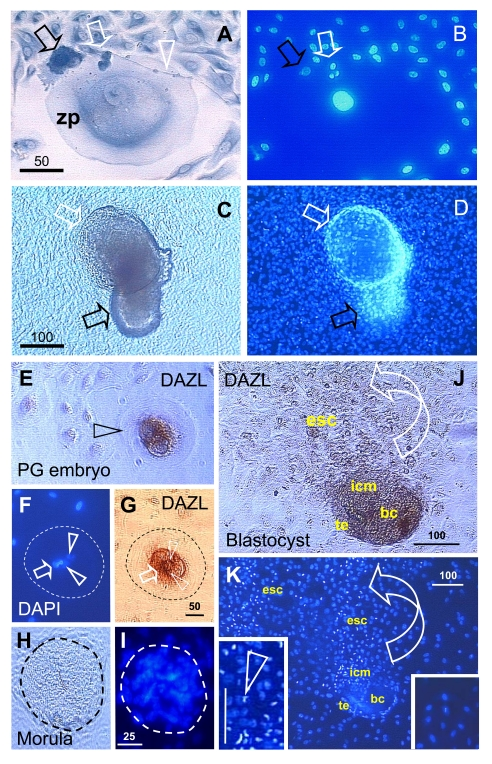
Figure 11. Oocyte and parthenote
development in vitro. (A) The oocyte development in OSC
culture is accompanied by a satellite (black arrow) and neuronal (white
arrow) cells. White arrowhead indicates neuronal extension. (B) DAPI
staining of (A). (C) The parthenote shows a blastocoele
(white arrow) and inner cell mass (black arrow). (D) DAPI staining
of (C). Four cell embryo. (E - G) and morula (H and I).
J and K panels show a blastocyst consisting of blastocoele
(bc), trophectoderm (te), and inner cell mass (icm) releasing ESC (esc).
Left insert in panel K shows enhanced DAPI staining of dividing ESC
vs. low DAPI staining of other cells in the culture (right insert). Details
in text. Adapted in part from Ref. [137], ©
Cambridge Journals.