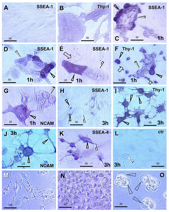
Figure 12. Human ovarian epithelial stem
cell cultures (representative images from four experiments): untreated (A and B); pre-treated for 1 day with E2 and 1h after TP+PG treatment (C-G);
pre-treated for 1 day with E2 and 3h after TS+PG treatment (H-O).
Lack of SSEA-1 expression (A) and moderate Thy-1 expression by some
epithelial cells (B). SSEA-1 is strongly expressed in some small
cells resembling stem cells (black vs. white arrowheads, C and D)
and one of the cells originating by asymmetric division (E). Similar
cells show strong expression of Thy-1 (F). The NCAM expression was
also detected in some cells (G). Two hours later, the cells reached
neuronal morphology and exhibited SSEA-1 expression in the cell bodies but
not extending processes (H, black vs. white arrowheads), Thy-1 and
NCAM expression in both (I and J, black arrowheads), and
SSEA-4 expression slightly exceeding that of SSEA-1 (K vs. H).
No staining was observed in the immunohistochemistry control (L).
Panels M-O show phase contrast microscopy with neuronal and
epithelial cells (M), floating numerous putative NSC (N), and
putative NSC exhibiting bubble type anchors (arrowheads, O). Numbers
above bars indicate microns. For details see text. Adapted from Ref. [138], © Landes Bioscience.