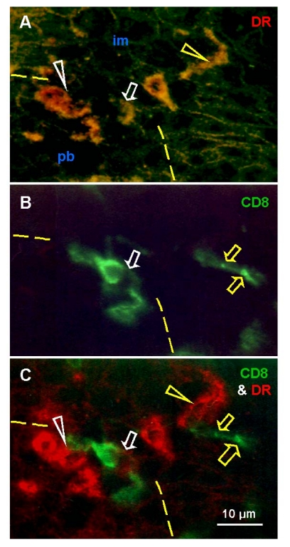
Figure 4.
Uterine cervix dual color
immunohistochemistry (HLA-DR peroxidase/CD8 FITC) viewed in dark field
visible light (A), incident fluorescence (B) and dark field
fluorescence (C). (A) Interface (dashed line) between
parabasal and intermediate layers. White arrowhead shows differentiating
DC, yellow arrowhead shows mature DC. Arrow indicates activated T cell with
HLA-DR expression (see below). (B) White arrow indicates T cell
exhibiting unusual elongated shape at the interface. Yellow arrows indicate
residual CD8 expression in fragmented T cell among adjacent im epithelial
cells. (C) Activated T cell with HLA-DR expression (white arrow)
interacts with differentiating DC (white arrowhead). Mature DC (yellow
arrowhead) accompany T cell fragmentation (yellow arrows). Reprinted from
Ref. [4], © Antonin Bukovsky.