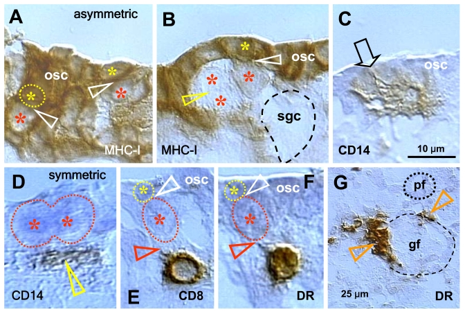
Figure 5.Expression of MHC class I heavy chain (MHC-I), CD14 of primitive MDC, CD8 of T cells, and HLA-DR (DR) of activated MDC and T cells, as indicated in the panels, in human fetal ovary obtained at midpregnancy (24 weeks). Asymmetric division (white
arrowheads, panels A and B) of OSC (osc) gives rise to the
OSC (yellow asterisks) and the germ cell daughters (red asterisks).
Symmetric division of germ cells follows (yellow arrowhead, panel B),
which is required for crossing over, and the secondary germ cells (sgc)
attain the ameboid shape (dashed line, no hematoxylin counterstain) to
leave the OSC layer and enter cortex. CD14+ primitive MDC interact with the
OSC (arrow, panel C) and accompany (arrowhead, panel D)
symmetric division of secondary germ cells. CD8 T cells (panel E)
and DR+ cells of lymphocyte type (panel F) accompany (red
arrowheads) asymmetric division of OSC (white arrowheads) resulting in
emergence of secondary germ cells. DR+ MDC (arrowheads, panel G)
associate with growing (gf) but not resting primordial follicles (pf). Bar
in C for A-F. Adapted from Ref. [36],
© Humana Press.