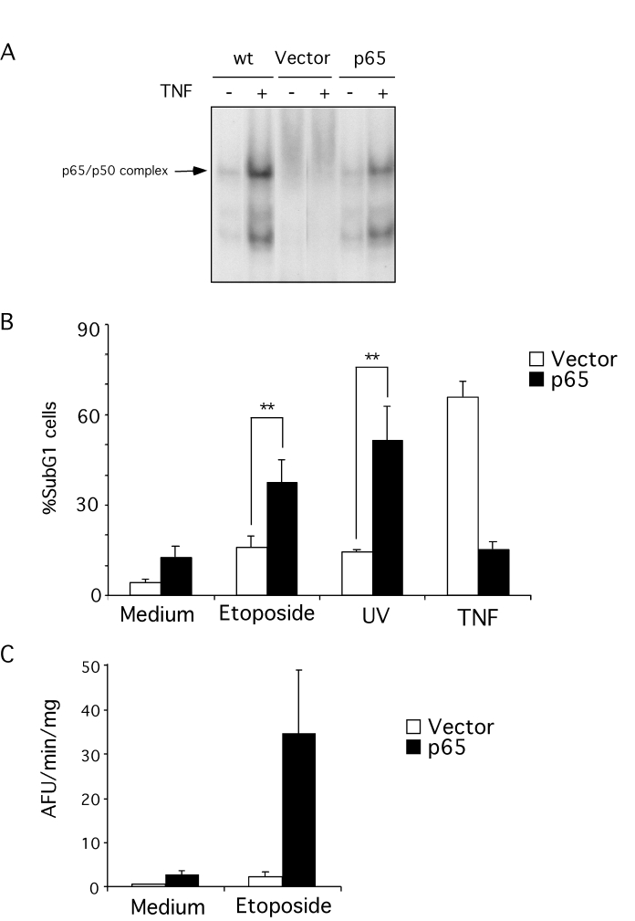
Figure 1.Apoptosis resistance in p65 null MEFs. (A)
retrovirus-mediated reconstitution of p65 null MEFs with p65 restores NF-κB function as measured by EMSA.
Wild type (wt), p65 null (vector) and p65 null reconstituted MEFs were
stimulated with 10 ng/ml TNFα for 6
hr. Nuclear proteins were extracted and equal amounts of extract incubated
with a radio-labeled NF-κB consensus probe. (B) p65 null
cells are resistant to genotoxin-induced apoptosis. Cells were treated with
10 μM etoposide or 5 mJ UV-irradiation for 18 hr. Floating and attached
cells were then collected and stained with propidium iodide (PI). DNA
content was analyzed by flow cytometry. Results are presented as percentage
of cells with sub-G1 DNA content. The data shown represent the mean and SEM of three
independent experiments. **statistically significant by student
t-test analysis (p<0.05). (C) S-100 extracts from p65 null
(vector) and reconstituted cells (p65) treated with 10 μM etoposide were used to assess caspase activity
by cleavage (arbitrary fluorescence units per minute [AFU/min]) of the
fluorogenic substrate, Ac-DEVD-afc. The data
shown represent the mean and SEM of three independent experiments.