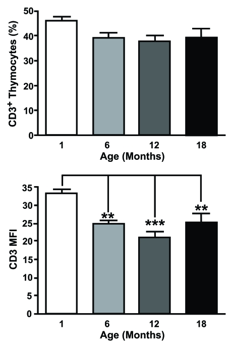
Figure 2.CD3 expression is altered on aged thymocytes. Thymocytes from different aged mice were stained with
anti-CD3 mAb and analysed by flow cytometry. The top histogram shows the percentage of CD3+ cells
positive and the bottom shows mean fluorescent intensity (MFI) of CD3 expression for one month old,
six month old, 12 month old and an 18 month old animals. MFI was obtained by gating on the entire population.
Although there were no age-related changes in the proportion of CD3+ thymocytes, a significant decrease
in the number of CD3 molecules on thymocytes associated with age was observed.
(One month n=5; six months n=5; 12 months n=8; 18 months n=4). **P<0.01; ***P<0.001.