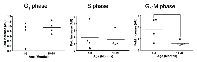
Figure 3.Cell cycle analysis on stimulated thymocytes from young and old mice. The various stages of the cell
cycle in thymocytes from young and old mice following treatment with ConA
and IL-2 after 24 hours was determined by flow cytometry. Data is expressed
as fold increase compared to time zero. It was observed that there was a
significant increase in the proportion of thymocytes from young mice at the
G2-M phase compared to thymocytes from older animals. One month
n=4; 18 months n=4. *P<0.05.