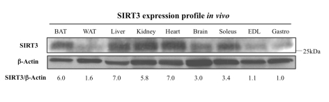
Figure 1.Tissue distribution of SIRT3 protein.
The SIRT3
protein is abundantly expressed in the brown adipose tissue (BAT), liver,
kidney, heart, brain, and soleus muscle, but very low in white adipose
tissue (WAT), the extensor digitorum longus muscle (EDL), or the
gastrocnemius muscle (Gastro). For each sample, 50 μg of protein was
loaded into a 10% acrylamide gel, electrophoresed, and transferred to a
nitrocellulose membrane. The membrane was probed using an anti-SIRT3 serum
or an anti-β-actin antibody. Blots were quantified with ImageQuant and
SIRT3/actin ratios are provided; since gastrocnemius (Gastro) has the
lowest SIRT3 expression in vivo, normalization (l.0) was set with
respect to this tissue.