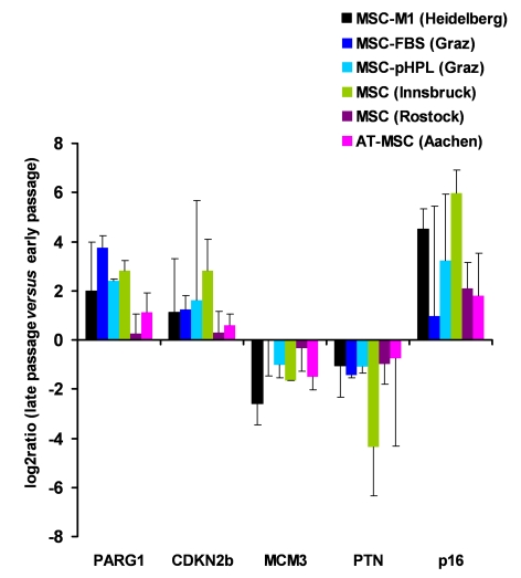
Figure 2.Gene expression markers for replicative senescence.MSC from human
bone marrow were either culture expanded as described before in medium-M1
with 2% fetal calf serum (M1, in Heidelberg, Germany [1]; n=3), in
culture medium with 10% fetal calf serum (FCS, n=2) or 10% pooled human
platelet lysate (pHPL, n=2; both in Graz, Austria
[38]),
in MEM supplemented with 20% FCS (Innsbruck, Austria [40];
n=2), and in MSCGM (Lonza) culture medium (Rostock, Germany; n=4).
Furthermore, MSC from adipose tissue were expanded with 10% pHPL (Aachen,
Germany,
n=4). RNA was isolated from corresponding early and late passages and
analyzed for differential gene expression in PARG1, CDKN2B, MCM3, PTN and p16ink4a. Primers and methods have
been described before [38]. These genes
did not facilitate reliable discrimination of senescent cells in all
samples but the tendency was consistent in all different MSC preparations.