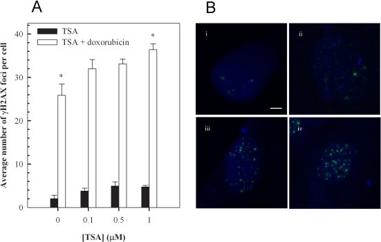
Figure 5.Trichostatin A (TSA) augments doxorubicin-induced accumulation of γH2AX foci in H9c2 cardiomyoctes. Cells pretreated with the indicated concentration of TSA for 24 hours were exposed to 1 μM doxorubicin for 1 hour, followed by a 24 hour treatment in fresh media. Cells were then stained for γH2AX foci, images were acquired with a Zeiss LSM 510 Meta Confocal microscope using 0.5 μm Z-sectioning and foci were quantitated using Metamorph (A). Mean ± standard deviations from two independent experiments (total of five independent experiments) are indicated (*p<0.001). Immunofluorescence visualization of γH2AX foci (B) in untreated H9c2 cells (i), cells treated with 1 μM TSA (ii), cells treated with 1 μM doxorubicin (iii) and cells treated with a combination of TSA and doxorubicin (iv) as described above. Bar = 2 μm; x 63 magnification.