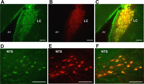
Figure 1.Dual immunofluorescence histochemistry for nesfatin-1 and tyrosine hydroxylase (TH).(A-C) Neurons immunoreactive (IR) to nesfatin-1 (green) (A) and TH (red) (B), and their merged image (C) in the LC. (D-F) Neurons IR to nesfatin-1 (green) (D) and TH (red) (E), and their merged image (F) in the NTS. Colocalization of nesfatin-1 with TH is seen by yellow color in (C) and (F). Scale bar, 100 μm. 4V, forth ventricle.