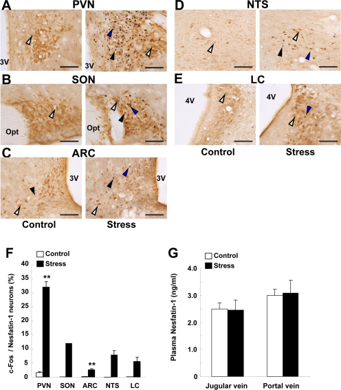
Figure 2.c-Fos expressions on nesfatin-1 neurons in several brain areas and plasma nesfatin-1 levels after restraint stress.(A-E) Double-immunohistochemical staining of c-Fos (black) and nesfatin-1 (brown) in the PVN (A), SON (B), ARC (C), NTS (D) and LC (E) in control conditions (left panels) and after restraint stress (right panels). White arrows indicate nefatin-1-IR neurons, black arrows c-Fos-IR neurons, and blue arrows both-IR neurons. (F) Incidence of c-Fos-IR neurons in nesfatin-1-IR neurons, as expressed by percentage. Number of c-Fos was significantly greater in stress than control conditions in the PVN and ARC, and found only in stress conditions in SON, NTS and LC. **p < 0.01 vs. control. (G) Nesfatin-1 concentrations in the plasma of jugular vein and portal vein were not different between control and stress conditions.