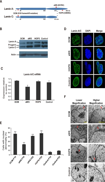
Figure 1.iPSCs from fibroblasts with 3 different mutations in lamin A/C(A) Simplified scheme depicting lamin A/C proteins and the corresponding amino acid (AA) substitutions (indicated with arrows) in DCM, aWS, and HGPS patients. (B) Western blot showing reduced expression of lamin A/C and accumulation of progerin in primary fibroblasts from DCM and HGPS patients respectively. Normal human fibroblasts were used as control. P indicates passage. Actin was used as loading control. A representative experiment is shown (this also applies hereafter when not mentioned otherwise). (C) qPCR analysis shows rather similar lamin A/C expression in primary fibroblasts from the 3 diseases compared to control fibroblasts. (D) Immunofluorescence photographs showing abnormalities of the nuclear membrane in primary fibroblasts from the 3 diseases compared to control fibroblasts. Nuclei are shown in blue. Scale bar indicates 10 μm. (E) Quantification of cells displaying abnormal nuclear membrane assessed by immunofluorescence microscopy and cell counting. The mean of 3 independent experiments +/− standard deviation (SD) is shown. ** indicates p value <0.01 measured with Student's t-test. (F) Electron microscopy photographs show nuclear membrane abnormalities in more detail. Arrows point to areas with more striking defects. Scale bars indicate 1 μm.