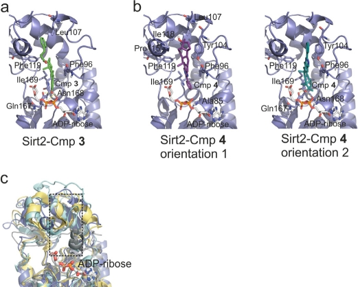
Figure 3.Models for the complexes between Sirt2/ADP-ribose and compounds 3 and 4, respectively(a) Docking model for the complex between the modeled Sirt2/ADP-ribose complex and 3. Residues forming the binding pocket proposed to be occupied by the compound are shown in stick presentation and labeled. (b) Docking models for the complex between the modeled Sirt2/ADP-ribose complex and 4. Two orientations representing poses about equally favored by the docking program are shown. Residues suggested to be involved in binding interactions are shown as sticks and labeled. (c) Overlay of the four Sirtuins studied here. Sirt2 is colored blue, Sirt3 yellow, Sirt5 cyan, and Sirt6 grey. Only the ADP-ribose of the Sirt2 complex is shown (sticks). The pocket suggested to bind 3 and 4 is indicated by a dotted box.