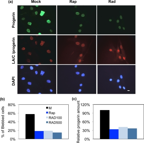
Figure 2.The nuclear morphology and progerin levels of rapamycin and RAD001 treated HGPS cells(a) The phenotype of nuclear blebbing was improved in RAD001 and rapamycin treated HGPS fibroblast cells. Cells were stained with DAPI (blue), laminA/C antibody (red), and progerin antibody (green) to show nuclear location and morphology. The treatment duration is for seven weeks. Mock: vehicle (DMSO, 0.025% v/v); Rap: 0.68 μM rapamycin, Rad: 0.1 μM RAD001. (Scale bar: 10 μm) (b) Quantification of the percentage of blebbing in all treatments. At least 200 nuclei were counted blindly. M: vehicle (DMSO, 0.025% v/v); Rap: 0.68 μM rapamycin; RAD100: 0.1 μM RAD001 treatment; RAD500: 0.5 μM RAD001 treatment. (c) Progerin was decreased in rapamycin or RAD001 treated HGPS fibroblasts. The relative amount of progerin was quantified using quantitative western blotting analysis and compared to the mock-treated HGPS cells.