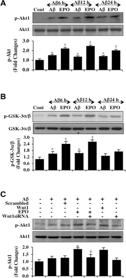
Figure 2.EPO maintains Akt1 activation through Wnt1 during Aβ exposure(A) Microglial protein extracts (50 μg/lane) were immunoblotted with phosphorylated Akt1 (p-Akt1) (active form) at 6, 12, and 24 hours following administration of Aβ (10 μM). Aβ resulted a mild increase in the expression of p-Akt1 over a 24 hour period. EPO (10 ng/ml) with a 1 hour pretreatment significantly increased the expression of p-Akt1 over a 24 hour period following Aβ exposure (*P < 0.01 vs. Control; †P< 0.01 vs. Aβ of corresponding time point). In all cases, each data point represents the mean and SEM from 3 experiments. (B) Akt1 activity in microglia was determined with a GSK-3β fusion protein through assessment of p-GSK-3α/β expression following Aβ exposure. EPO (10 ng/ml) with a 1 hour pretreatment significantly increased the activity of Akt1 over 12 hours following Aβ exposure (*P <0.01 vs. Control; †P<0.01 vs. Aβ of corresponding time point). In all cases, each data point represents the mean and SEM from 3 experiments. (C) Gene reduction of Wnt1 was performed with transfection of Wnt1 siRNA prior to Aβ exposure in microglia and p-Akt1 expression was determined at 6 hours following Aβ exposure. Loss of Wnt1 resulted in a decreased expression of p-Akt1 following a 6 hour period of Aβ exposure and significantly reduced EPO (10 ng/ml) expression of p-Akt1 during Aβ exposure. Non-specific scrambled siRNA did not alter p-Akt1 expression during Aβ exposure (*P < 0.01 vs. Aβ; †P < 0.01 vs. EPO/Aβ).