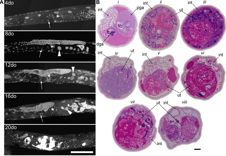
Figure 1.Germline masses accumulate with agea, Partial maximum projection of mid-section of whole wild-type worms stained with DAPI at 4, 8, 12, 16, and 20-days-old raised at 20 degrees. Arrow indicates the -1 oocyte in diakinesis (none visible in 20-day-old worm). Asterisk indicates spermatheca. Large arrowhead indicates unfertilized, endomitotic nuclei. # indicates a mass that has no distinct cellular structure. Small arrowhead in the 20-day-old indicates site of invasion from uterus to spermatheca. Scale bar represents 100 microns. b, Cross sections of a 20-day-old wild-type worm stained with pararosaniline and methylene blue. Cross sections from the same worm starting at approximately -1 oocyte position along anterior half (i). Remaining sections are spaced 50 μm apart (ii-viii), moving towards posterior and ending at approximately -1 oocyte position along posterior half (viii). In some cross sections, the uterine growth has taken up nearly the whole diameter of the worm (ii, iii, vii). Growth recedes at midbody (iv). Intestine (int), distal gonad arm (dga), proximal gonad arm (pga), and uterus (ut) are indicated.