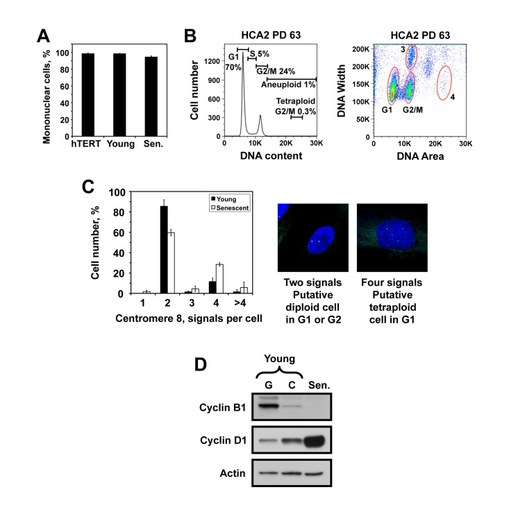
Figure 2.The senescent cell population with 4N DNA content is not due to polynucleated cells or tetraploid cells.(A) The number of nuclei per cell was counted in hTERT-immortalized, young, and senescent HCA2 cells after staining with DAPI and tubulin. (B) PI staining and flow cytometric analysis of pre-senescent HCA2 cells showing the absence of tetraploid G2 population. The left panel is the conventional DNA content histogram, and the right panel is a corresponding area versus width plot. Zone 3 contains two-cell aggregates of cells with 2N DNA content, and zone 4 shows a putative position of tetraploid G2 cells with 8N DNA content. (C) Fluorescent in situ hybridization with a probe to chromosome 8 centromeric region in young and senescent HCA2 cells. Diploid G1 or G2 cells show two signals, while tetraploid G1 cells are expected to show four signals. Hypothetically, if sister chromatids separate after a prolonged G2 arrest a diploid G2 cells may also show four signals. The percentages of cells with indicated numbers of chromosome 8 centromeric signals are plotted. The experiment was repeated three times and error bars show s.d. (D) Cyclin levels in replicatively senescent cells. Western blot of total cell lysates from HCA2 cells probed with cyclin B1 (Abcam, ab72-100) and cyclin D1 (Abcam, ab10540-100) antibodies.