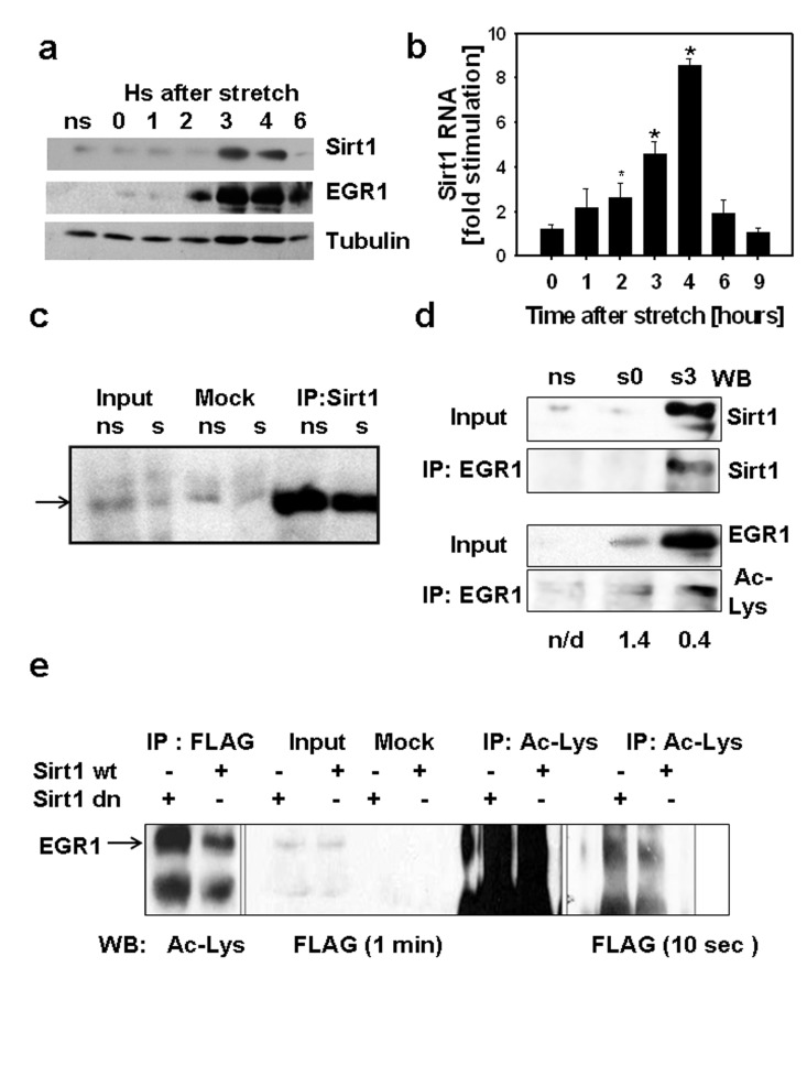
Figure 1.(a) C2C12 myotubes were cultured on 7 FlexCell plates, 6 plates were subjected to 30 min stretch while a set of myotubes was kept without stretch (ns), stretched myotubes were harvested at different times after stretch from 0 to 6 hours. 80 μg of total protein from each sample and molecular weight markers (MW) were subjected to SDS-PAGE and Western blot with antibodies against SIRT1, EGR1 and tubulin. Molecular weight markers positions are displayed on the left of the western blot scan images. (b) SIRT1 RNA content was determined by RT-real time PCR in stretched and non-stretched myotubes by RT-real time PCR and fold stimulation represents the stretched/non-stretched SIRT1 RNA content ratios (*indicates statistical significant difference from non-stretched) (c) Stably expressing FLAG-EGR1C2C12 cells (C2C12 12.4 clone) were plated in flexible bottomed plates and a set of cells were stretched (s) whereas another set was used as non-stretched controls (ns). Total proteins were obtained 3 hours after stretch; 0.5 mg of protein were incubated with protein A/G agarose beads loaded with a rabbit SIRT1 antibody or without antibody (mock); 50μg of total protein (input) and mock and SIRT1 immunoprecipitates were subjected to SDS-PAGE and Western blot with a monoclonal anti-FLAG M2 peroxidase conjugated antibody (Sigma-Aldrich). (d) Total proteins (0.4 mg) from non-stretched myotubes (ns), myotubes subjected to 30 min stretch and harvested immediately (s0) or 3 hours after (s3) were immunoprecipitated with anti-EGR1, 75 μg of total protein (input) and immunoprecipitates from each sample were analyzed by Western blot with a mouse monoclonal anti-SIRT1 (Sigma-Aldrich) or a mouse monoclonal anti-acetyl lysine (Upstate). The numbers below each lane represent the estimate ratio of acetylated EGR1.total EGR1 obtained by densitometric analysis of the films. (e) 0.75 mg of total protein from C2C12 12.4 cells transfected with pYE-Sir2 or pYE-Sir2 (H/Y) were incubated overnight without (mock) or with a rabbit polyclonal anti-acetyl lysine antibody and immunoprecipitated with A/G agarose beads; 80ug of total protein and total immunoprecipitates were subjected to Western blot with anti-FLAG M2 peroxidase, images of the film exposed for 1 min (right) or 10 sec(left) are shown.