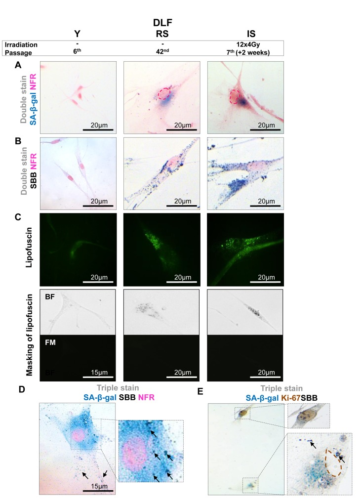
Figure 1.Lipofuscin accumulates and co-localizes with Senescence-Associated beta-galactosidase (SA-β-gal) in sub-confluent senescent primary human diploid lung fibroblasts (DLF)Y (Young): Early- passage cells, RS: Replicative-senescent cells and IS (irradiated): Early passage cells that became prematurely senescent after irradiation (12×4Gy). Collected cells were fixed on slides with 4% parafolmadehyde (A) All three cultures were stained with SA-β-gal and nuclear fast red as counterstain (NFR). Cells from RS and IS cultures acquired the characteristic senescent morphological phenotype (enlarged and flattened) and were positive for SA-β-gal staining (turquoise color). (B) All cultures were stained with Sudan Black B (SBB) and NFR. Cells from RS and IS cultures, which had the morphological phenotype of senescence, were also positive for SBB (dark blue-black granules). (C) Top panels: green pseudocolor represents visualization of lipofuscin's autofluorescence at 450-490 nm. Bottom panels: RS and IS cells that stained with SBB (BF, bright field microscopy) show no auto-fluorescence of lipofuscin (FM, fluorescence microscopy without pseudocolor), indicating that SBB stains lipofuscin. Cells with the morphological phenotype of senescence were positive for both SA-β-gal and SBB (D), while cells that were positive for Ki67 were negative for both SA-β-gal and SBB (E). Insets: Cells at higher magnification, pink dashed lines: indicate NFR-stained nuclei, brown dashed lines: indicate Ki67- negative nuclei, black arrows: show SBB granules.