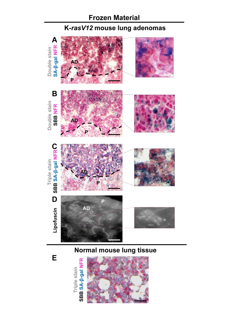
Figure 5.Lipofuscin and Senescence-Associated beta-galactosidase (SA-β-gal) activity co-localize in lung adenomas demonstrating senescence in a mouse model conditionally expressing K-rasV12 in the lungFrozen sections derived from mouse lung K-rasV12 adenomas. (A) Cells from the adenomas show SA-β-gal activity. (B) Characteristic perinuclear deposition of blue black granules in cells stained with Sudan Black B (SBB), representing positivity for lipofuscin. (C) Cells from lung adenomas positive for both, SA-β-gal activity and lipofuscin. (D) Fluorescence microscopy (at 450-490 nm) verifying lipofuscin presence. (E) Normal mouse lung tissue negative for SA-β-gal activity and lipofuscin. P: Parenchyma, AD: Adenoma. Scale bars: A-C, 200 μm; D, 25 μm; E, 50 μm. Insets: Cells at higher magnification.