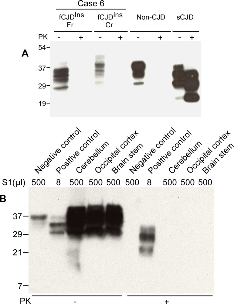
Figure 2.Detection of PK-sensitive PrPSc. (A) Conventional Western blotting of PrP treated with or without PK in case 6. Fr: frontal cortex; Cr: cerebellum. No PrP was observed after PK treatment in the samples from both fCJDIns (case 6) and non-CJD. The PK-resistant PrP27-30 was indicated in the sample from sCJD. The migration of PrP from the cerebellum of case 6 was slightly slower than that of PrP from both non-CJD and sCJD controls. (B) Precipitation of abnormal PrP by NaPTA. S1 from non-CJD (500 μl), sCJD (8 μl), and case 6 (three brain regions: 500 μl each) was incubated with NaPTA and then was subjected to SDS-PAGE and immunoblotting with 3F4. Although a small amount of PrP was precipitated from non-CJD brain sample (500 μl of S1), no PK-resistant PrP fragments were detected. NaPTA was able to precipitate PrP from 8 μl of sCJD S1 (62.5-fold less than non-CJD S1) and the precipitated PrP was resistant to PK-digestion. Compared to non-CJD sample, NaPTA precipitated large amounts of PrP from three different brain regions of case 6 including the cerebellum (Cr), occipital cortex (Oc) and brain stem (BS). After PK-treatment of the NaPTA-precipitated PrP from case 6, no PrP bands were observed.