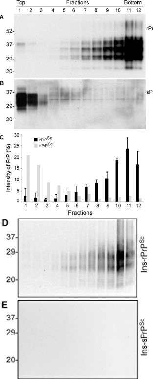
Figure 4.Comparison of oligomeric state of PrP in case 6 and other cases with rPrPSc by sucrose step gradient sedimentation. (A) Western blotting of PrP in individual fractions of sucrose gradient analysis of brain homogenate from case 3 with readily detectable rPrPSc. (B) Western blotting of PrP in individual fractions of sucrose gradient analysis of brain homogenate from case 6 with sPrPSc. C: Bar graph of PrP in individual fractions from three 144-bp insertion mutation cases with rPrPSc (average of PrP percentages from the three fCJDIns+rPrPSc cases) and case 6 with no rPrPSc. Blots were probed with 3F4 antibodies. D and E: PrP in individual fractions from cases 3 (D) and 6 (E) was detected by Western blotting after treatment with PK at 0.5 μg/ml. PrP was only detected in fCJDIns+rPrPSc but not in fCJDIns+sPrPSc.