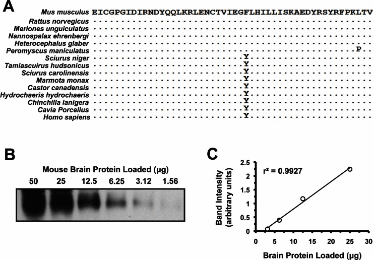
Figure 1.Quantitative analysis of IGF1R protein levels. (A) Alignment of the 50 amino acids comprising the epitope recognized by the anti-IGF1R polyclonal antibody used (SC-712, see Experimental Procedures). This region maps to the N-terminus of the protein, which is on the alpha-chain peptide of IGF1R. It excludes the signaling peptide, which is cleaved during protein maturation, and was not used in the immunogen. Human sequence is included due to it being used to generate the antibody. Sequence identity is indicated by dots. (B) Western blot of serial dilutions of mouse brain whole tissue protein extracts. Linear region was between 25 and 3.12 μg. (C) Band intensity in arbitrary units, plotted against protein loaded to verify linearity.