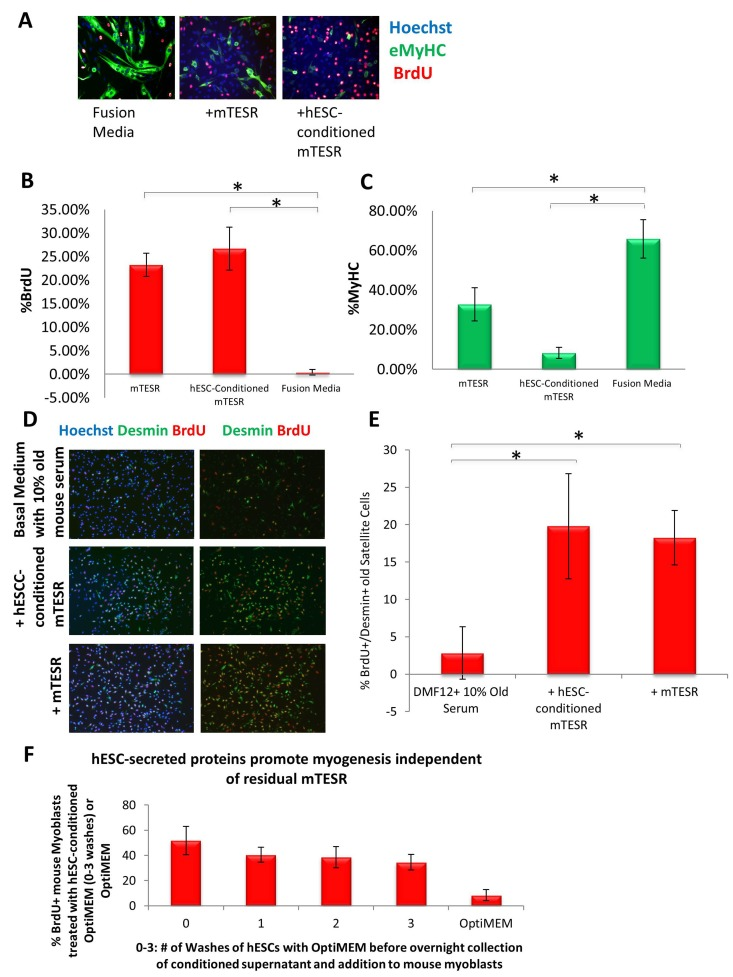
Figure 1.Both mTeSR-1 and hESC-Conditioned mTeSR-1 increase primary myoblast and satellite cell Proliferation and inhibit Differentiation(A) Primary Mouse Myoblasts were cultured for 24 hours in 50% fusion/differentiation medium (DMEM, 2% horse serum) plus 50% of the specified medium. A 2 hour BrdU pulse was performed before cell fixation to label proliferating cells. Immunofluorescence was performed for eMyHC (green) and BrdU (red), with Hoechst (blue) labeling all nuclei. Representative images are shown. Proliferation and differentiation of fusion-competent myoblasts were quantified by cell scoring in 25 random fields of each condition using a Molecular Devices automated imager and MetaXpress cell scoring software. Results are displayed as the mean percent of BrdU+ (B) or eMyHC+ (C) proliferating or differentiating cells +/−SD, respectively. N=4 *P< 4×10⁻¹⁰ for BrdU+ myoblasts -incubated in 50% mTeSR-1 as compared to myoblasts incubated in just fusion medium, or 50% hESC-conditioned mTeSR-1 as compared to myoblasts incubated in just fusion medium. *P< 0.005 for eMyHC+ fusing myoblasts in 50% mTeSR-1 as compared to myo-blasts incubated in fusion medium alone, and *P< 9×10⁻⁵ for eMyHC+ fusing myoblasts in 50% hESC-conditioned mTeSR-1 as compared to myoblasts incubated in just fusion medium. (D) Old injury activated myofiber-associated satellite cells were isolated at 3 days post cardiotoxin-induced muscle injury, and cultured overnight in 50% DMEM/F12 with 10% old serum, and 50% of the medium specified, followed by a 2 hour BrdU pulse to label proliferating cells before cell fixation. Immunofluorescence was performed with Desmin (green) and BrdU (red), with Hoechst (blue) labeling all cell nuclei. Representative images are shown and demonstrate that both hESCconditioned mTeSR-1 and mTeSR-1 have a pro-myogenic effect on activated satellite cells. (E) Proliferating Desmin+/BrdU+ satellite cells were quantified by cell scoring in multiple random fields of each condition. Results are displayed as the mean percent of BrdU+/Desmin+ proliferating satellite cell cells +/−SD. N=3, *P< 0.05 for satellite cells in 50% mTeSR-1 as compared to satellite cells incubated in just basal medium with old serum, and *P< 0.001 for satellite cells in 50% hESC-conditioned mTeSR-1 as compared to satellite cells incubated in just basal medium with old serum. (F) Undifferentiated hESCs that were grown in mTeSR-1 medium were washed 0-3 times with Opti-MEM, followed by overnight incubation in Opti-MEM and collection of the resulting conditioned Opti-MEM. The hESC-conditioned Opti-MEM was spun down to remove cell debris, before addition to myoblasts as a 50/50 mix with myogenic fusion medium for culture overnight. A 2 hour BrdU pulse was performed to label proliferating cells prior to cell fixation and immunofluorescence was performed with eMyHC and BrdU, with Hoechst labeling all cell nuclei (images not shown). Proliferating and differentiating cells were quantified by cell scoring 25 random fields of each condition using an automated imager and MetaXpress cell scoring software. Results are displayed as the mean percent of BrdU+ or eMyHC+ proliferating or differentiating cells +/−SD, respectively. N=2