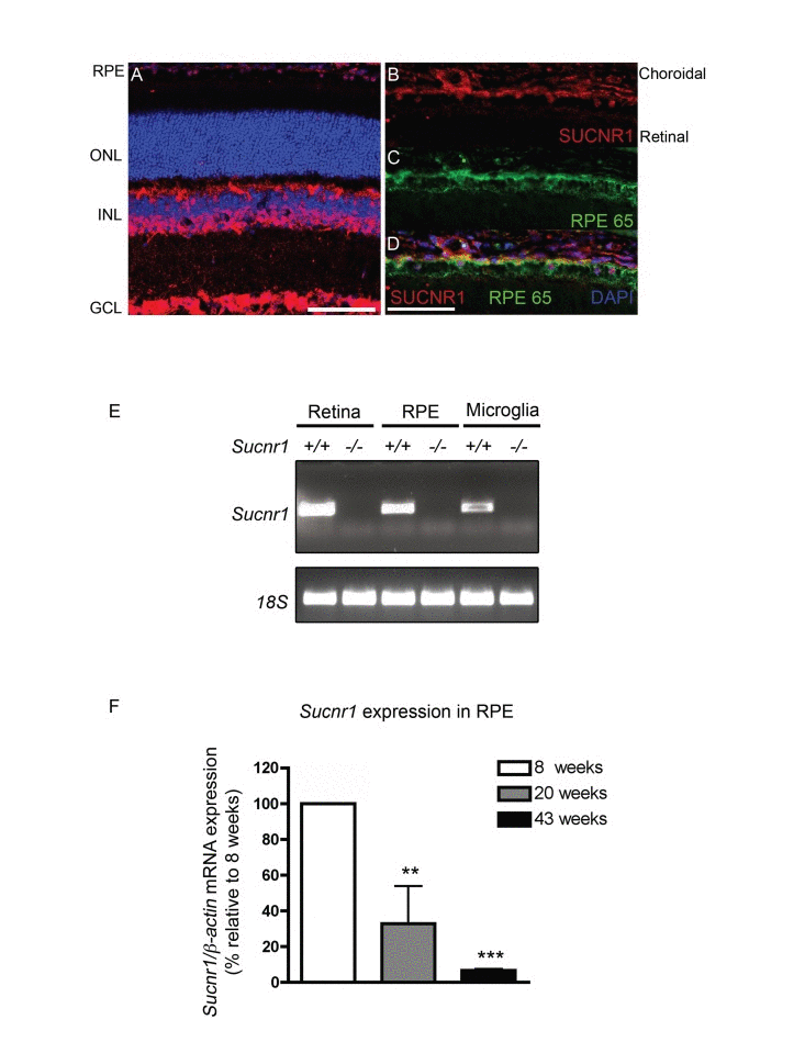
Figure 1.SUCNR1 is expressed in RPE and microglia and diminishes with age(A) Immunohistochemistry on sagittal retinal cryosections reveals expression of SUCNR1 in the GCL, INL and RPE. (B-D) Confocal imaging corroborates expression of SUCNR1 (red) in the RPE as confirmed by co-localization with the RPE marker RPE65 (green). Images are representative of 3-4 experiments. (E) Expression profile of Sucnr1 mRNA shows transcripts in RPE and CNS microglia from wild-type mice while an absence of transcripts is noted in samples from Sucnr1−/− mice. (F) Quantitative PCR on RPE isolated from 8, 20 or 43 week old wild-type mice shows a steady decrease of Sucnr1 levels with age (n=4-6). Values are expressed as percentage of controls ± S.E.M., normalized to β-actin standards. ONL: outer nuclear layer, INL: inner nuclear layer, GCL: ganglion cell layer and RPE: retinal pigment epithelium. Scale bars (A-D): 100μm.