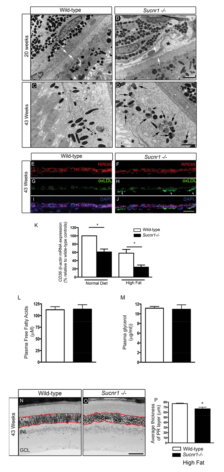
Figure 2.Deficiency in SUCNR1 leads to outer retinal lesionsTransmission electron microscopy of RPE/sub-retina the in wild-type (A) and Sucnr1−/− (B) at 20 weeks of age reveals regional disruption of BM and presence nodular debris (asterix). By 43 weeks of age, lipofuscin granules (black arrows) accumulate in the sub-retina of Sucnr1−/− mice (D) while minimal lipofuscin is detected in wild-type mice. Confocal microscopy on retinal cross sections demonstrates accumulation of sub-retinal deposits of oxLDL in Sucnr1−/− mice (F,H,J) while wild-type controls remained ox-LDL free (E,G,I). Images are representative of 3 distinct experiments. (K) Quantitative PCR reveals lower levels of scavenger receptor CD36 in isolated RPE extracts in Sucnr1−/− mice when compared to wild-type controls (n=5-10). Values are expressed as percentage of controls ? S.E.M, normalized to ?-actin standards. *P<0,05. Similar levels of plasma FFA (L) and glycerol (M) were noted in both wild-type and Sucnr1−/− mice suggesting that systemic lipolysis was not affected by SUCNR1. (N-P) Toluidine blue-stained epoxy retinal semi-thin sections show mild degeneration of photoreceptors (20 points of analysis per retina; n=3-4 mice) *P<0,05. Scale bar (A-D): 2μm; (E-J): 100μm; (N,O): 75μm.