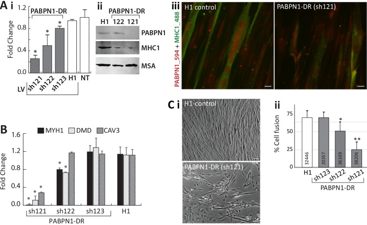
Figure 6.PABPN1-DR in human myotubes causes myogenic defectsHuman myotubes were transduced with shRNA specific to PABPN1 (sh121, sh122, or sh123) or H1 empty vector. Non-transduced (NT) cells were used as controls. (A) i Bar-chart shows PABPN1 mRNA expression in stably-transduced myoblasts. Fold change was normalized to GapDH housekeeping gene and to a non-transduced culture. Averages are of 6 biological replicates. ii Western blot analysis of PABPN1, MHC1 and muscle actin (MSA), as a loading control in sh121, sh122 or H1 myotube cultures. iii Immunofluorescence of PABPN1 (labelled with Alexa-594) and MHC1 (labelled with Alexa-488) in sh121 or H1 myotube cultures. Scale bar 10 μm. (B) Bar chart shows fold change of MHC1, DMD, and CAV3 in 121-, 122-, 123-, and H1- myoblast cultures. Fold change was normalized to GapDH and to a non-transduced culture. Averages are of 3 biological replicates. Significant down-regulation (P<0.05) is indicated with asterisks. (C) i- images of H1 controls and the PABPN1 down regulation sh121 fused cultures. Scale bar is 50 μm. ii- Chart bar shows the percentage of nuclei in fused myotubes that express MHC1 (cell fusion) in H1 sh123 sh122 or sh121 myotube cultures. Averages and SD are from six replicates and the number of nuclei that were quantified per sample is indicated within each bar. Significant effect in PABPN1-DR cultures from control cultures (P<0.05 or P<0.005) is indicated with one or two asterisks, respectively.