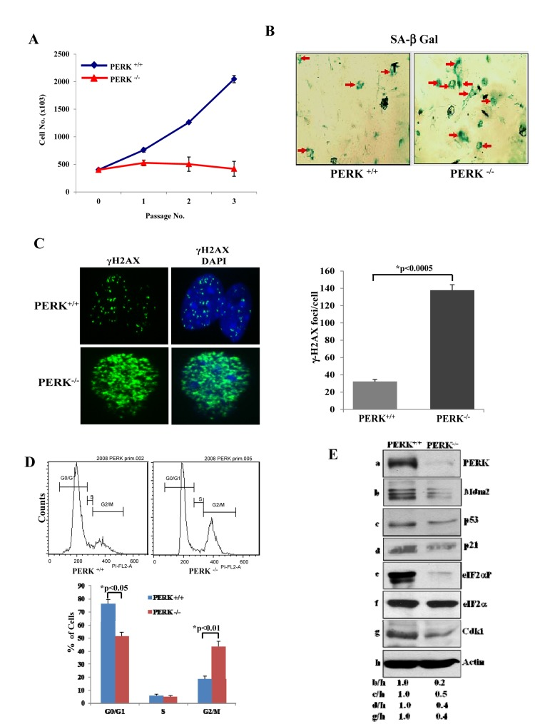
Figure 3.PERK deficiency impairs proliferation and induces premature senescence(A) Primary PERK+/+ and PERK−/− MEFs were maintained in culture for the indicated passages and their proliferation was assessed by cell counting. The data represent an average taken from two independent experiments performed in triplicates. (B) Induction of senescence was evaluated by SA β-Gal staining from cells in passage 3. Senescent cells are indicated by arrows. (C) DNA damage was assessed by γ-H2AX staining and fluorescence microscopy of cells in passage 3. Nuclei were visualized by DAPI staining. Histograms represent the average number of γ-H2AX foci per cell (n=100). (D) PERK+/+ and PERK−/− MEFs from passage 3 were subjected to PI staining and FACS analysis to determine cell cycle progression. Histograms show the percentages of cells in G0/G1, S and G2/M from three independent experiments. (E) Protein extracts (50 μg) from MEFs maintained in passage 3 were immunoblotted for the indicated proteins.