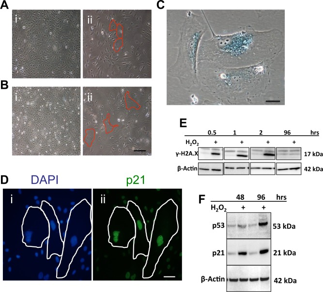
Figure 1.Oxidative stress induces senescence in ECs(A) HUVECs were treated with 0.2mM H2O2. After 2 days cells began to demonstrate the morphology of EC senescence (ii-Red outline) compared to untreated cells (i). (B) After a further 2 days the number of senescent cells had increased and the size of the senescent cells had also increased (ii-Red outline), compared to untreated cells (i). This is a representative of 10 HUVEC lines. Bar=220μm. (C) HUVECs were treated with 0.2 mM H2O2 and after 4 days stained for SA-β-gal. This is a representative of 10 HUVEC lines. Bar=25μm. (D) HUVECs were untreated (i) or treated with 0.2 mM H2O2 and after 4 days stained using immunofluorescence for DAPI (i) and p21 (ii). This is a representative of 5 HUVEC lines. Bar=50μM. (E) and (F) Cells were treated with 0.2 mM H2O2 and lysates analysed for levels of γH2A.X (E), p53 and p21 (F). β-Actin was used as a loading control. This is a representative of 8 HUVEC lines.