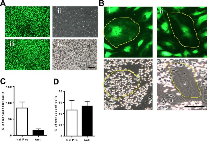
Figure 4.Oxidative stress induced senescence has a unique inflammatory phenotype(A) Normal HUVECs were stained with cell tracker green for visualization (i and iii) and then neutrophil adhesion was determined by a static adhesion assay. Cells were either untreated (i,ii) or treated with 5ng/ml of TNFα for 5 hours (iii, iv). (B) HUVECs were treated with 0.2 mM H2O2. After 4 days the cells were stimulated with 5ng/ml TNFα and stained with cell tracker green for visualization. Senescent cells are highlighted with a yellow line. Representative photos of the pro-inflammatory senescent cells (i) and the anti-inflammatory senescent cells (ii) are shown. This is a representative of 6 HUVEC lines. Bar=25μm. (C) From the photographs taken in (B) the number of anti- and pro-inflammatory senescent cells was determined and given as the % of total senescent cells. This is the representative of the mean +/− SD of 200 senescent cells from 6 HUVEC lines. (D) From videos taken of neutrophil rolling and adhesion (Videos 1-5) the percentage of pro and anti-inflammatory senescent cells after TNFα stimulation was determined. This is a representative of the mean +/− SD of 220 senescent cells from 4 HUVEC lines.
Figure 4 — Age-associated stresses induce an anti-inflammatory senescent phenotype in endothelial cells | Aging