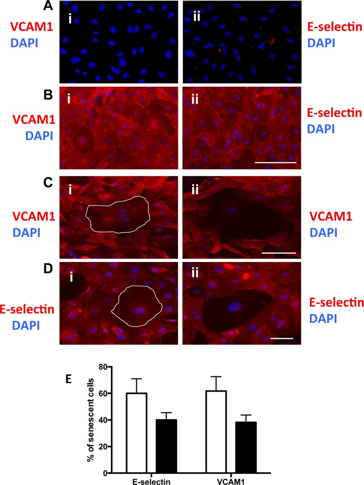
Figure 5.Senescent cells have altered adhesion molecule expression(A and B) HUVECs were either untreated (A) or stimulated with 5ng/ml TNFα (B) and stained for the surface expression of VCAM-1 (red) (i) and E-selectin (red) (ii) and co-stained with DAPI (blue). This is a representative of 6 HUVEC lines. Bar=100μm. (C) HUVECs were treated with 0.2 mM H2O2, for 4 days, stimulated with 5ng/ml TNFα then fixed and stained for the surface expression of VCAM-1 (red) and co-stained with DAPI (blue). Representative pro-inflammatory senescent cells (i) and anti-inflammatory senescent cells (ii) are shown, highlighted by the white outline. This is a representative of 6 HUVEC lines. Bar=100 μm. (D) Cells were treated as in (C) but stained for the surface expression of E-selectin (red) and co-stained with DAPI (blue). This is a representative of 6 HUVEC lines. Bar=100μm. (E) From the photographs taken in (C and D) the number of anti and pro inflammatory senescent cells was determined. This is a representative of the mean percentage +/− SD of 150 senescent cells from 6 HUVEC lines.