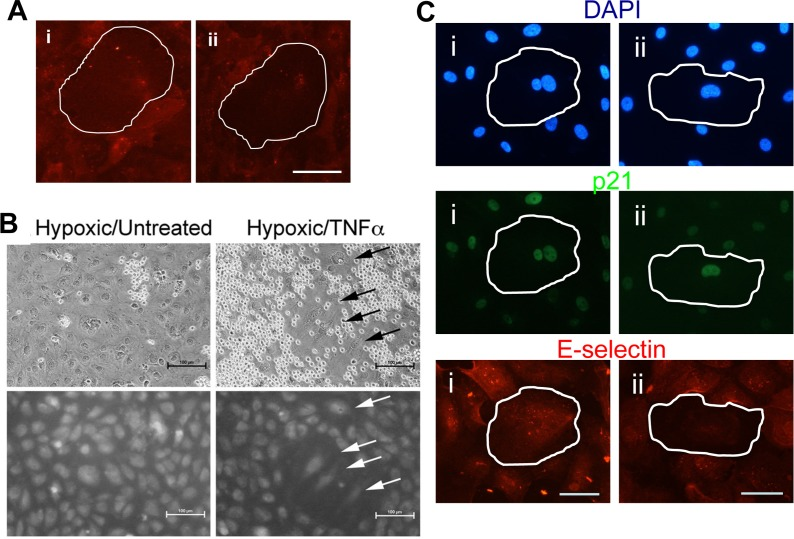
Figure 7.Shear stress and hypoxia induce an anti-inflammatory phenotype(A) HUVECs were subjected to 48hrs of flow at 2dyne/cm2. The cells were then treated with 5ng/ml of TNFα for 5 hours, fixed and stained for E-selectin (red). Induced pro-inflammatory senescent cells (i) and anti-inflammatory senescent cells (ii) are shown. A senescent cell is highlighted by the white outline. This is a representative of 3 HUVEC lines. Bar=100μm. (B) Binding of neutrophils to HUVECs that had been cultured at 0.5% oxygen for five days followed by treatment for six hours with 5ng/ml TNFα (left hand panel). Upper panels show light microscopy of neutrophils upon HUVECs. Lower panels show CMFDA-labelled HUVEC monolayers. Cells with a senescent morphology are indicated with an arrow. Hypoxic treated cells that were not stimulated with TNFα are shown in the left hand panel. Shown are representative images of three experiments using independent HUVEC lines. (C) HUVCs exposed to 10μM DFO for 72 hrs were treated with 5ng/ml of TNFα for 5 hours, fixed and stained for p21 (green), E-selectin (red) and co-stained with DAPI (blue). Representative pro-inflammatory senescent cells (i) and anti-inflammatory senescent cells (ii) are shown, highlighted by the white outline. This is a representative of 3 HUVEC lines. Bar=50μm.