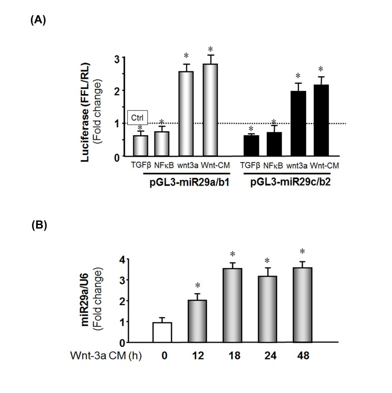
Figure 5.Wnt-3a induces miR-29 promoter activation(A) MPCs were transfected with pGL3-miR-29a/b1 (gray bar) or pGL3-miR-29C/b2 (black bar) to assay miR-29 promoter activities. Transfected cells were treated as follows: left to right, treated with TGF-β; co-transfected with NFκB plasmid (pCMV1.p65); transduced with Ad-Wnt-3a; or treated with Wnt-3a-conditioned media. The bar graph represents firefly luciferase activity corrected for renilla luciferase activity (FFL/RL) and compared to control (luciferase activity of pGL3 untreated, set to 1 and designated by a horizontal line in the graph). The data represent the means ± s.e.; (n=9, *p<0.05 vs. pGL3 (Ctrl). (B) MPCs were cultured in Wnt-3a conditioned media and cells harvested at the indicated times. Total RNA was isolated and miR-29a expression was measured by qPCR. The bar graph shows miR-29a from Wnt-3a conditional medium expressed as a fold-change vs. miR-29a levels in cells cultured with control media (set to 1). Results are normalized to U6 RNA as an internal control. The data represent the means ± s.e.; (n=3 pairs; *p<0.05 vs. control).