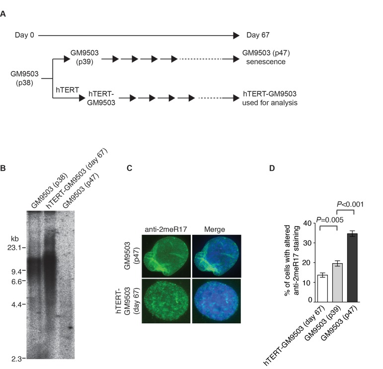
Figure 6.Introduction of hTERT into normal primary fibroblast cells suppresses the formation of senesence-associated altered nuclear staining of methylated TRF2. (A) Schematic diagram of the experimental setup. At day 0, GM9503 cells were infected with retrovirus expressing hTERT, generating hTERT-GM9503 cells. Both GM9503 and hTERT-GM9503 cells were cultured continuously for 67 days. (B) Genomic blots of telomeric restriction fragments from GM9503 (p38), GM9503 (p47) and hTERT-GM9503 at day 67. About 3 μg of RsaI/HinfI-digested genomic DNA from each sample was used for gel electrophoresis. The DNA molecular size markers are shown to the left of the blots. (C) Analysis of indirect immunofluorescence with anti-TRF2-2meR17 antibody. Cell nuclei of GM9503 and hTERT-GM9503 were stained with DAPI in blue. (D) Quantification of percentage of cells with altered nuclear staining of methylated TRF2. At least 900 cells in triplicate were scored in blind for each cell line as indicated. Standard deviations from three independent experiments are indicated.