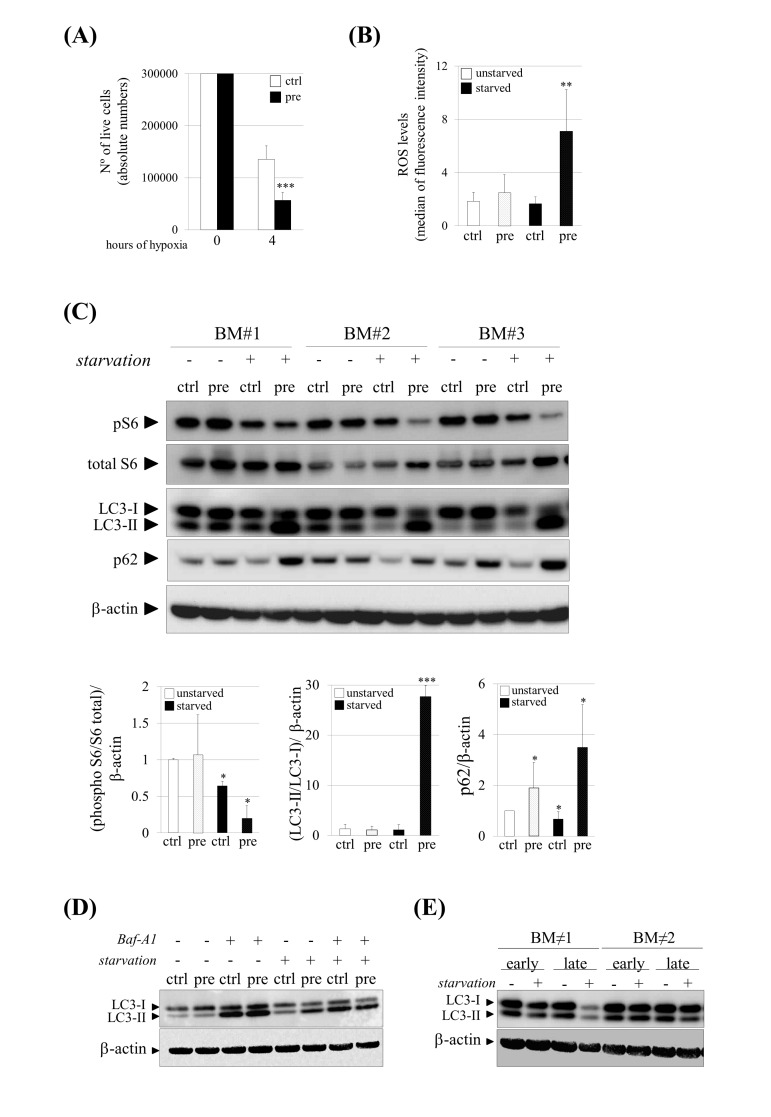
Figure 2.Increased susceptibility and impaired autophagy under stress conditions in pre-hMSCs. (A) Number of live cells after submitting hMSCs to hypoxia. (B) Reactive Oxygen Species (ROS) measurement of hMSCs cultured under basal (unstarved) or starvation conditions (starved). (C) Western blot of indicated proteins in ctrl and pre-hMSCs cultured under basal or serum starvation conditions. β-actin was used as loading control. hMSCs from three independent bone marrow donors (BM) where analyzed together. Densitometry for each immunoblot is provided. (D) Determination of autophagic flux of hMSCs treated with Bafilomycin (Baf-A1) 0.1 μM during 9 hours. LC3-I and LC3-II expression levels are shown and β-actin was used as loading control. (E) Western blot of LC3-I and LC3-II expression levels in replicative senescent hMSCs (Early: early-passage hMSCs; Late: late-passage hMSCs), Bars are average +/- standard deviation of 3 independent donors. Differences marked with asterisks are significantly different from control unstarved (panel A and D) or control starved cells (panel B) *** p<0.001, ** p<0.01, *p<0.05.
(pre):prelamin A-accumulating hMSCs, (ctrl):control-hMSCs.