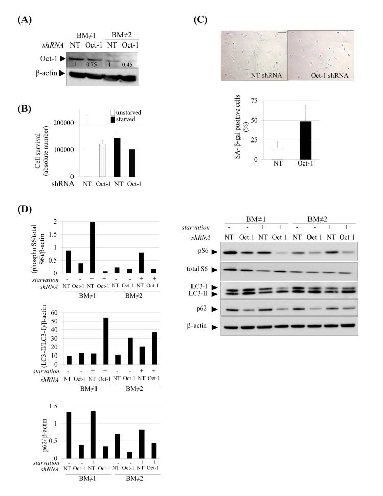
Figure 6.Induction of senescence and autophagy in Oct-1 silenced hMSCs. (A) Western blot showing decreased Oct-1 expression after Oct-1 shRNA plasmids transduction in hMSCs, NT: Non Targeting shRNA. Numbers show densitometric quantification for Oct-1 expression. (B) Cell survival after Oct-1 silencing in hMSCs cultured under basal (unstarved) or serum starvation conditions (starved). (C) Representative SA β-gal staining (left) and quantification (right) in hMSCs transduced with NT or Oct-1 shRNA. Scale bar: 100 μm. (D) Western blot of indicated proteins from hMSCs silenced for Oct-1 or not (NT). β-actin was used as loading control. Densitometry for each immunoblot is provided.
For these experiments hMSCs from two independent donors were used(BM≠1 and BM≠2). Bars are mean +/- standard deviation