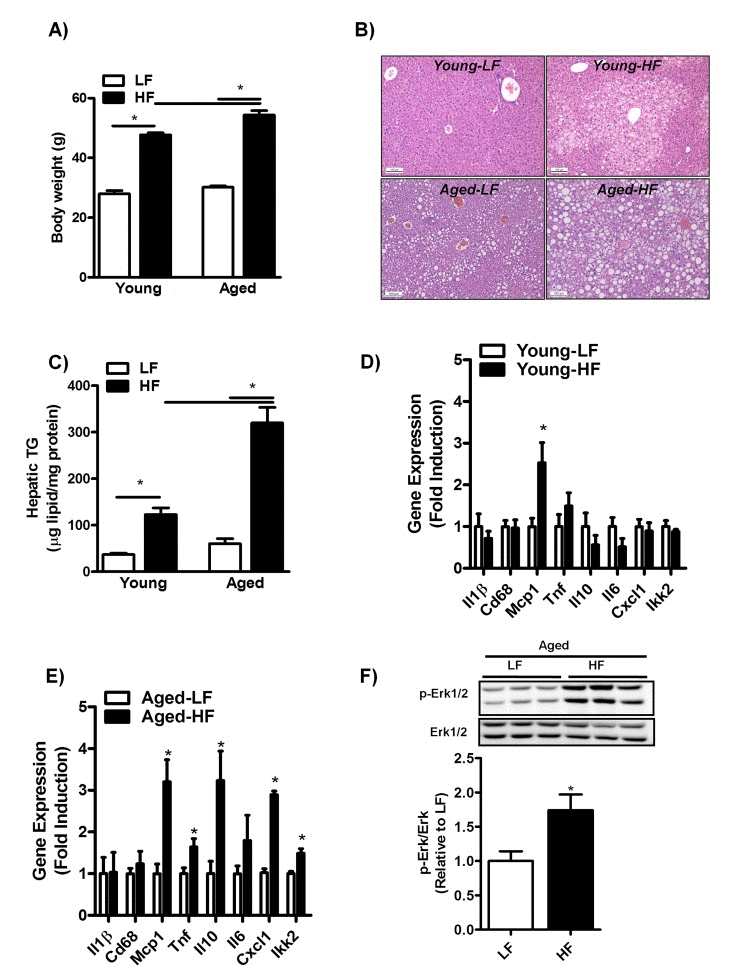
Figure 3.Aging increases hepatic steatosis and inflammation in mice fed a HFD for 12 weeks(A) Body weight of young and middle-aged mice fed a HFD. (B) H&E staining of paraffin embedded liver sections and (C) biochemically quantification of liver triglycerides (TG) of young and middle-aged mice fed a HFD. (D) mRNA expression of cytokines and genes involved in inflammation interleukin-1 (Il1), Cluster of Differentiation 68 (Cd68), monocyte chemoattractant protein-1 (Mcp-1), tumor necrosis factors (Tnf), interleukin-10 (Il10), interleukin-6 (Il6), Chemokine (C-X-C motif) ligand 1 (Cxcl1), IkappaB kinase 2 (Ikk2) in the liver of young mice fed a HFD (versus LFD-fed mice) for 12 weeks determined by qRT-PCR and expressed as fold induction. (E) mRNA expression of cytokines and genes involved in inflammation Il1,Cd68, Mcp-1, Tnf, Il10, Il6, Cxcl1, Ikk2 in the liver of middle-aged mice fed a HFD. (F) Immunoblot analysis using anti-Phospho-Erk1/2 and anti-Erk1/2 antibody was performed in liver extracts of middle-aged mice fed a LFD or HFD for 12 weeks, tubulin antibody was performed as a control for protein loading (not shown). Values are expressed as mean ±SEM; n = 6-8 mice in each group. *p ≤0.05 (nonparametric Mann-Whitney U test).