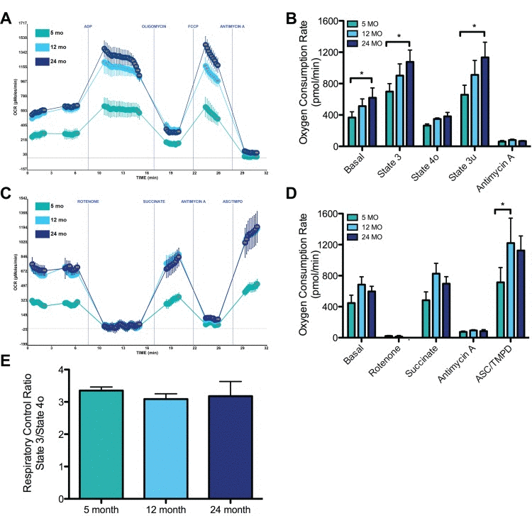
Figure 4.Effects of aging on synaptic mitochondrial bioenergeticsSynaptic mitochondria from 5, 12, and 24 month mice were isolated and assayed as described in the Materials and Methods. (A) Representative graph output of coupling assay of isolated synaptic mitochondria. Point-to-point oxygen consumption rate (OCR) data are shown with succinate as the substrate followed by addition of ADP, oligomycin, FCCP, and antimycin A. (B) Basal (complex II), state 3, state 4o, state 3u respiration. *Significantly (p<0.05) lower in 5 vs. 24 month old animals, n = 5. (C) Representative graph output of electron flow assay. Point-to-point OCR data are shown with pyruvate and malate as the substrate followed by the addition of rotenone, succinate, antimycin A, and ASC/TMPD. (D) OCR response following rotenone inhibition of complex I, succinate driven complex II, antimycin A inhibition of complex III, and ASC/TMPD driven complex IV. *Significantly (p<0.05) lower in 5 vs. 12 month old animals, n = 3. (E) Respiratory Control Ratio (State 3/State 4o) shows no change between 5, 12, and 24 months.