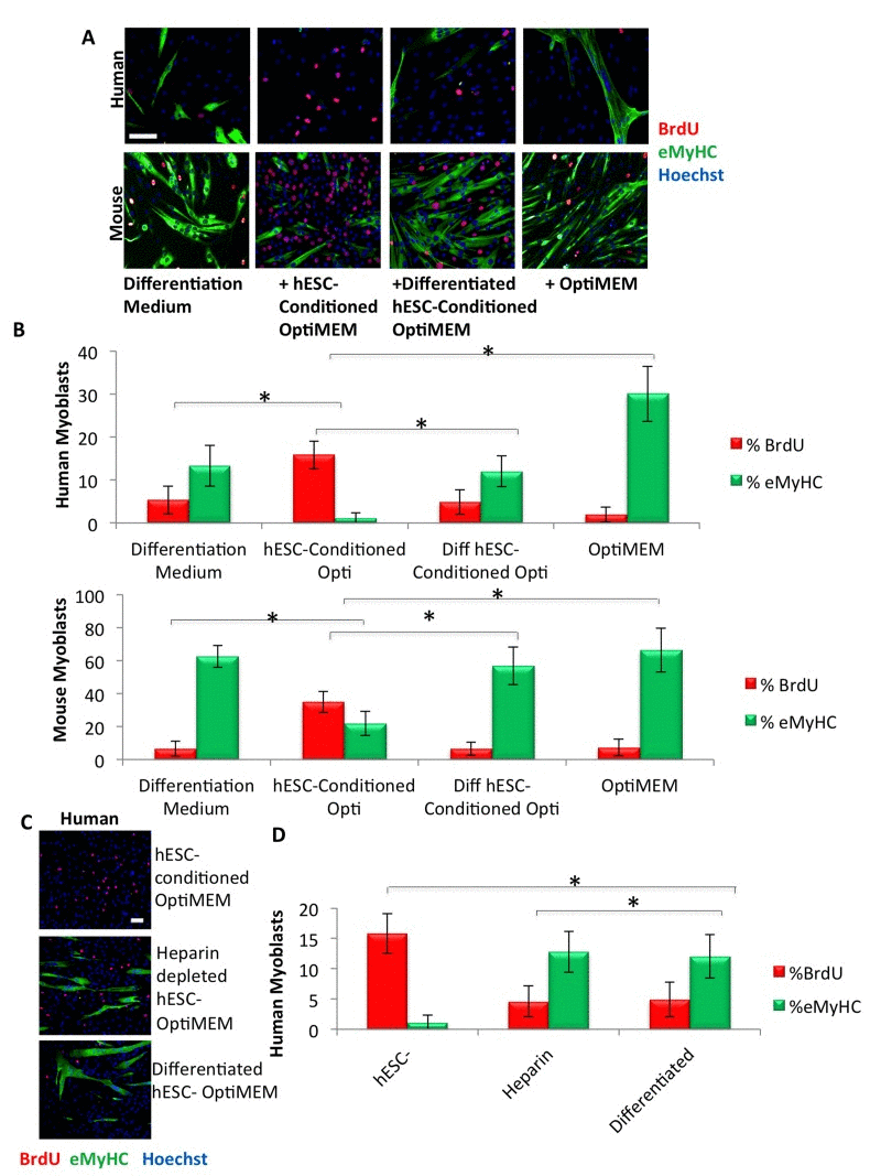
Figure 1.Pro-regenerative embryonic factors that enhance human and mouse myoblast proliferation contain heparin binding domains(A) Primary human or mouse myoblasts were cultured for 24 hours or 72 hours, respectively, in 50% differentiation medium (DMEM, 2% horse serum) plus 50% of the specified medium, with daily medium changes. A 4 hour or 2 hour BrdU pulse on human or mouse myoblasts, respectively, was performed before cell fixation to label proliferating cells. Immunofluorescence was performed for eMyHC (green) and BrdU (red), with Hoechst (blue) labeling all nuclei. Representative images are shown. Scale bar = 100 μM (B) Proliferation and differentiation of fusion-competent myoblasts were quantified by cell scoring in 25-50 random fields of each condition using a Molecular Devices MetaXpress automated imager and cell scoring software. Results are displayed as the mean percent of BrdU+ or eMyHC+ proliferating or differentiating cells +/−SD, respectively (n=6). Significant differences were identified by Student's t-tests (*p<0.004 for human cells; *p<8×10⁻¹⁹ for mouse cells). (C) Primary human myoblasts were cultured, BrdU pulsed, immunostained and quantified as in (A), with the specified medium. Representative images are shown. Scale bar = 100 μM (D) Proliferation and differentiation of fusion-competent human myoblasts were quantified as in (B). Results are displayed as the mean percent of BrdU+ or eMyHC+ proliferating or differentiating cells +/−SD, respectively (n=6). Significant differences were identified by Student's t-tests (*p<0.004).