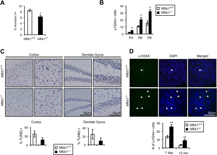
Figure 4.Loss of Nfkb1 is associated with decreased apoptosis and increased γH2AX accumulation(A) Annexin V binding in primary Nfkb1+/+ and Nfkb1−/− MEFs at passage 2 (n=5 MEF isolates per group, harvested at different times). (B) γH2AX staining in primary Nfkb1+/+ and Nfkb1−/− MEFs at the indicated passage. MEF data show mean +/− SD. (C) Percentage of cells that are TUNEL-positive in the cortex and dentate gyrus in 12-month old Nfkb1+/+ and Nfkb1−/− mice (n=4 animals per group). Representative sections shown (above). (D) Total number of γH2AX foci in brain sections from newborn and 12-month old Nfkb1+/+ and Nfkb1−/− mice (n=3 animals per group). γH2AX positive cells were calculated as described in the methods, data represent mean +/− SEM. Representative coronal brain sections demonstrate co-localization of γH2AX and DAPI (arrow heads). *p<0.05. **p<0.01.