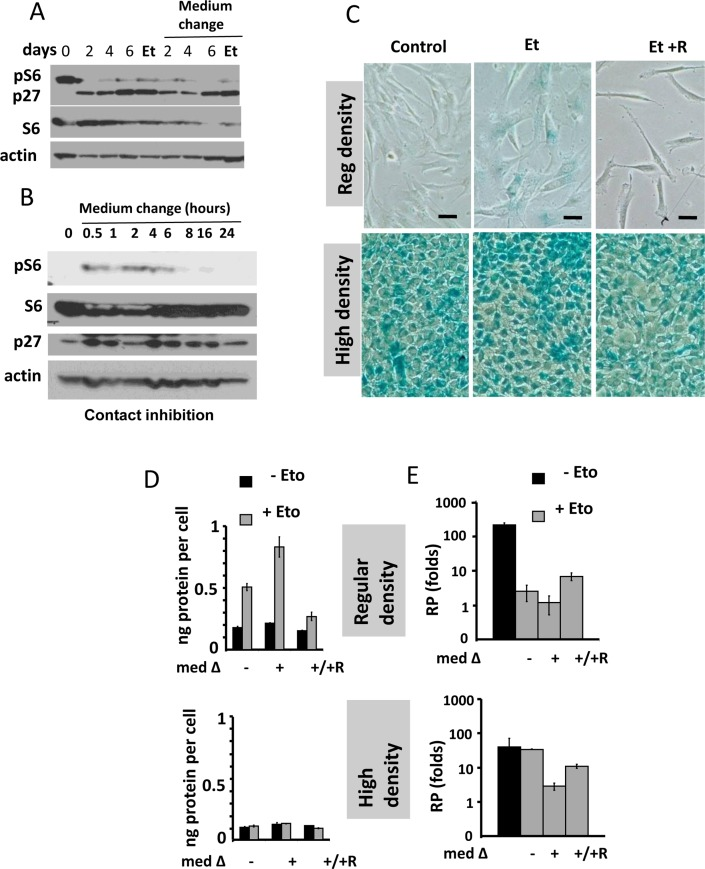
Figure 1.Contact inhibition suppresses etoposide-induced senescence in WI-38t cells(A-B) Immunoblot analysis [41]. (A) WI-38t cells [41] were plated at high density and lysed on the days indicated. When indicated “Medium change”, the medium was changed to fresh one every day. Et: cells were treated with 0.5 μg/ml etoposide on day 3 and lysed on day 6. p-S6(S240/244). (B) The effect of medium change on Contact Inhibited cells, measured in hours. (C) Beta-gal staining. WI38t cells were plated at regular or high density. After 3 days, 0.5 μg/ml etoposide (Et) and +/− 10 nM rapamycin (R) was added, if indicated. After 3 days, cells were stained for beta-Gal. Bar – 100 μm. (D-E) Cells were treated as described in panel C. Data are mean ± SD. (D) Cell size, protein per cell. (E) Reversibility potential or Replicative potential (RP). On day 6, cells were counted and re-plated at 1000/well in 12-well plates in fresh drug-free medium. Cells were counted after 9 days of growth. Fold increase in cell numbers were calculated. Mean ± SD.
Figure 1 — Gerosuppression in confluent cells | Aging