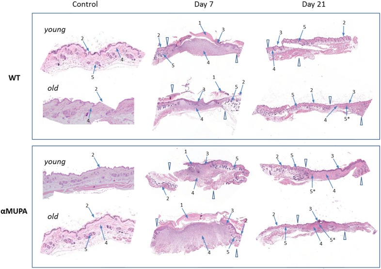
Figure 3.Histological examination of skin wound healing in αMUPA and WT miceHematoxylin and eosin [H&E] staining of the skin samples of Day 0 (control), Day 7 (early re-epithelialization) and Day 21 (full wound closure) after operation. Magnification: 4x. Arrowheads indicate the wound edges. Arrows: 1 – granulation tissues; 2 – normal epidermis; 3 – hyperplastic epidermis; 4 – fibroblasts; 5 – hair follicles; 5* – new hair follicle formation within re-epithelialized edges.