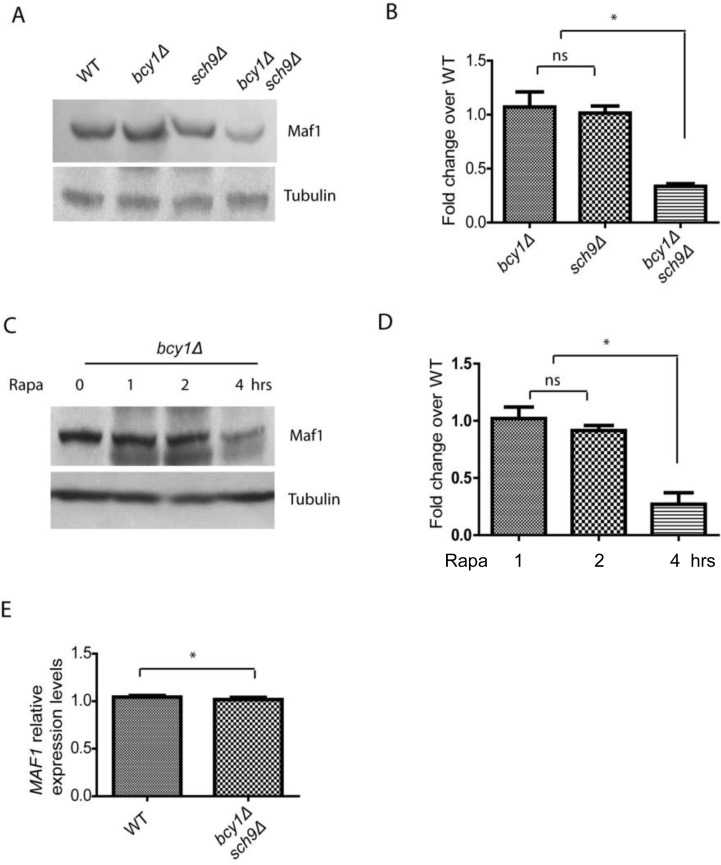
Figure 2.PKA and Sch9 coordinate to control Maf1 protein levels(A) Maf1 protein levels are reduced by PKA hyper-activation in sch9Δ cells. WT, bcy1Δ, sch9Δ and sch9Δ bcy1Δ cells expressing a Myc-tagged Maf1 (Maf1-Myc) from a low copy centromeric plasmid were cultured to early log phase and protein samples were prepared for western blot analysis. Protein amount were controlled by endogenous tubulin levels. (B) Quantification of Maf1 protein levels from 2 independent experiments as shown in A. ns, not significant; * p < 0.01. (C) Maf1 protein levels are reduced by PKA hyper-activation in growth inhibiting conditions. bcy1Δ cells expressing a Myc-tagged Maf1 (Maf1-Myc) from a low copy centromeric plasmid were treated with 100 nM rapamycin for 1, 2, 4 hours and protein samples were prepared for western blot analysis. Protein amount were controlled by endogenous tubulin levels. (D) Quantification of Maf1 protein levels from 2 independent experiments as shown in C. ns, not significant; * p < 0.01. (E) MAF1 mRNA levels are not reduced by sch9Δ bcy1Δ as shown by real-time qPCR.