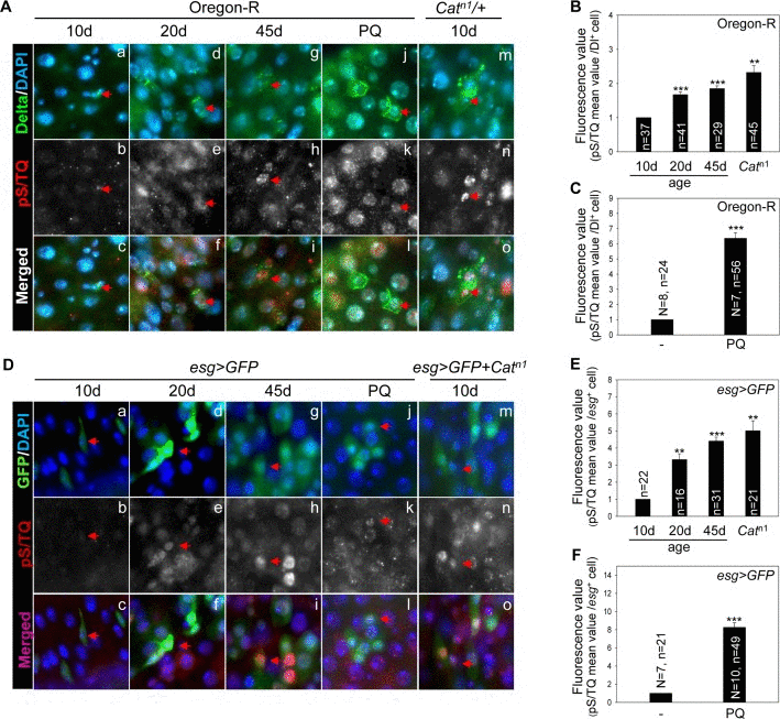
Figure 1.The pS/TQ signal increases with age and under the influence of oxidative stress in Drosophila intestinal stem cells (ISCs)(A) The age- and oxidative stress-induced increase in pS/TQ signals in Delta protein-positive (Dl+) small cells. (a–i) An age-related increase of pS/TQ signals in Dl+ small cells. The gut of 10- (a–c), 20- (d–f), and 45-day-old (g–i) wild-type flies was stained with anti-Dl (green) and anti-pS/TQ (red) antibodies and 4′,6-diamidino-2-phenylindole (DAPI; blue). (j–o) An oxidative stress-induced increase in pS/TQ signals in Dl+ small cells. The gut of 10-day-old 10 mM PQ-fed (j–l) and Catn1/+ (m–o) flies was labeled with anti-Dl (green) and anti-pS/TQ (red) antibodies and DAPI (blue). Red arrows indicate Dl+ small cells. c, f, i, l, and o are merged images. The original magnification is 400×. (B–C) The florescence value of pS/TQ signals in Dl+ small cells as a function of age and oxidative stress. The gut of 10-, 20-, and 45-day-old wild-type flies and 10-day-old Catn1/+ (B) and 10 mM PQ-fed (C) flies was labeled with anti-GFP (green) and anti-pS/TQ (red) antibodies and DAPI (blue). The fluorescence intensity of pS/TQ signals in the Dl+ cells was measured in the 5a and b regions of the posterior midgut. The variable n indicates the number of Dl+ cells. Fluorescence intensity of the cells, which are exactly in focus, was measured in approximately 10 to 14 midgut specimens; ***p < 0.0001, **p < 0.001. (D) An age- and oxidative stress-induced increase in pS/TQ signals in esg+ small cells. (a–i) The age-related increase in pS/TQ signals in esg+ small cells. The gut of 10- (a–c), 20- (d–f), and 45-day-old (g–i) esg>GFP flies was labeled with anti-GFP (green) and anti-pS/TQ (red) antibodies and DAPI (blue). (j–o) The oxidative stress-induced increase in pS/TQ signals in esg+ small cells. The gut of 10-day-old esg>GFP 10 mM PQ-fed (j–l) and esg>GFP+Catn1 (m–o) flies was labeled with anti-GFP (green) and anti-pS/TQ (red) antibodies and DAPI (blue). Red arrows indicate esg+ cells. c, f, i, l, and o are merged images. The original magnification is 400×. (E–F) The fluorescence values of pS/TQ signals in esg+ small cells as a function of age and oxidative stress. The gut of 10-, 20-, and 45-day-old esg>GFP and 10-day-old esg>GFP+Catn1 (E) and 10 mM PQ-fed (F) flies was labeled with anti-GFP (green) and anti-pS/TQ (red) antibodies and DAPI (blue). The fluorescence intensity of pS/TQ signals in esg+ cells was measured in the 5a and b regions of the posterior midgut. The variable n indicates the number of esg+ cells; ***p < 0.0001, **p < 0.001.