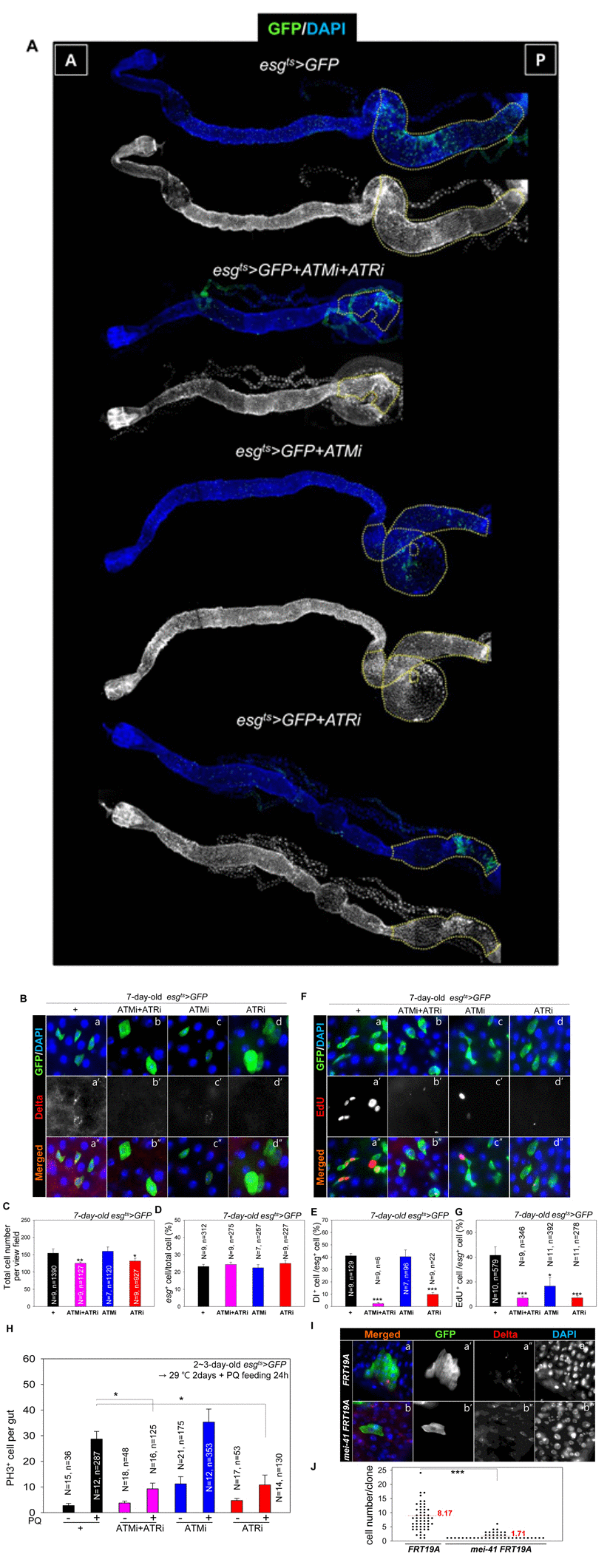
Figure 5.ATM and ATR are required for proliferation and maintenance of Drosophila intestinal stem cells (ISCs)(A) The phenotype of the midgut with an ISC/enteroblast (EB)-specific knockdown of ATM, ATR, or both. The gut specimens of esgts>GFP, esgts>GFP+ATMi+ATRi, esgts>GFP+ATMi, or esgts>GFP+ATRi flies (kept at 29 °C for 14 days) were labeled with an anti-GFP (green) antibody and 4′,6-diamidino-2-phenylindole (DAPI, blue). The yellow dots indicate the posterior region. The regions with yellow dots show a large esg-GFP+ population in a particular region. The original magnification is 100×. (B) A loss of Delta protein-positive (Dl+) cells in the midgut with an ISC/EB-specific knockdown of ATM, ATR, or both. The gut specimens of esgts>GFP (a-a''), esgts>GFP+ATMi+ATRi (b-b''), esgts>GFP+ATMi (c-c''), or esgts>GFP+ATRi flies (d-d'') (kept at 29 °C for 7 days) were labeled with anti-GFP (green) and anti-Dl (red) antibodies and DAPI (blue). a''–d'' are merged images. The original magnification is 400×. (C) A graph showing the total cell number in the gut with an ISC/EB-specific knockdown of ATM, ATR, or both. N: the number of gut specimens, n: the total cell number. *p < 0.01. **p < 0.001. (D) A graph showing the the ratio of esg+ cells to total cells in the midgut with an ISC/EB-specific knockdown of ATM, ATR, or both. N: the number of gut specimens, N: the number of gut specimens, n: the esg+ cell number. (E) A graph showing the ratio of Dl+ cells to esg+ cells in the midgut with an ISC/EB-specific knockdown of ATM and ATR. N: the number of gut specimens, n: the Dl+ cell number. ***p < 0.0001. The numbers of cells of each cell type were counted in the R5 region of the posterior midgut under a microscope. (F) The gut specimens of esgts>GFP (a), esgts>GFP+ATMi+ATRi (b), esgts>GFP+ATMi (c), or esgts>GFP+ATRi flies (d) (kept at 29 °C for 7 days with subsequent feeding on EdU-containing media for 24 h) were stained with anti-GFP (green) and anti-EdU (red) antibodies and DAPI (blue). The original magnification is 400×. (G) A graph showing the ratio of EdU+ cells to esg+ cells in the midgut with an ISC/EB-specific knockdown of ATM, ATR, or both. The numbers of cells of each cell type were counted in the R5 region of the midgut. N: the number of gut specimens, n: the esg+ cell number. *p < 0.01. ***p < 0.0001. (H) Effects of the ISC/EB-specific knockdown of ATM, ATR, or both on the oxidative stress-induced increase in proliferation of ISCs. The gut specimens of esgts>GFP (black), esgts>GFP+ATMi+ATRi (pink), esgts>GFP+ATMi (blue), and esgts>GFP+ATRi (red) flies (kept at sub 29 °C for 2 days, with subsequent feeding on media containing 10 mM PQ for 24 h) were labeled with anti-PH3 (red) and anti-GFP (green) antibodies and DAPI (blue). The numbers of PH3+ cells were counted in the whole midgut. The data (mean ± SE) from esgts>GFP (PQ-/+), esgts>GFP+ATMi+ATRi (PQ-/+), esgts>GFP+ATMi (PQ-/+), and esgts>GFP+ATRi (PQ-/+) flies were collated from 15, 12, 18, 16, 21, 12, 17, and 14 gut specimens, respectively. N: the number of gut specimens, n: the mitotic cell number; *p < 0.01. (I) Effect of mei-41 null mutant MARCM on ISC maintenance of posterior midguts. The flies carrying hsFLP, tubP-GAL80, neoFRT 19A/neoFRT 19A; UAS-mCD8::GFP/+; tubP-GAL4/+ (WT) or hsFLP, tubP-GAL80, neoFRT 19A/mei-41[G0221b] neoFRT 19A; UAS-mCD8::GFP/+; tubP-GAL4/+ (mei-41 null mutant) were dissected and marked with anti-Dl (red), anti-GFP (green) antibodies and DAPI (blue) at 7 days after induction. The original magnification is 400×. (J) A graph showing the clone size of WT and mei-41 mutant clone. The numbers of cells of each clone were counted in the posterior region of 9-10 midguts. ***p < 0.0001.