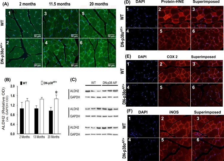
Figure 7.(A-C) Expression levels of ALDH2, a juvenile protective factor, by the gastrocnemius of young (2 mos), middle aged (13.5 mos) and aged (20 mos) WT and DN-p38αAF/+ mice(A: upper panel) The levels of expression of ALDH2 in the gastrocnemius of WT mice. (A1) young (2.0 mos); (A2) middle aged (13.5 mos); (A3) aged (20 mos) mice. (A: lower panel) The levels of expression of ALDH2 by the gastrocnemius of DN-p38αAF/+ mice; (A4) young (2.0 mos); (A5) middle aged (13.5 mos), and (A6) aged (20 mos) mice. (B) Bar graph presentation of the western blot analyses of the data in (C) presented as the relative OD vs. β-actin levels. *p < 0.05 vs corresponding WT. (C) Western immunoblot analyses of the levels of ALDH2 in young (2.0 mos), middle aged (13.5 mos) and aged (20 mos) WT and DN-p38αAF/+ mice. (GM, right leg). *p = 0.05 vs. WT of the same age. (D-F) Expression levels of protein-HNE complexes, COX2, and iNOS by young (3 mos) WT and DN-p38αAF/+ mice. (D) The level of expression of protein-HNE in the gastrocnemius of young (3mos) WT mice DN-p38αAF/+ mice (D4-6). (E) The level of expression of COX2 in the gastrocnemius of young (3mos) WT mice (E1-3) and DN-p38αAF/+ mice (E4-6). (F) The level of expression of iNOS in the gastrocnemius of young (3mos) WT (F1-3) and DN-p38αAF/+(F4-6) mice.