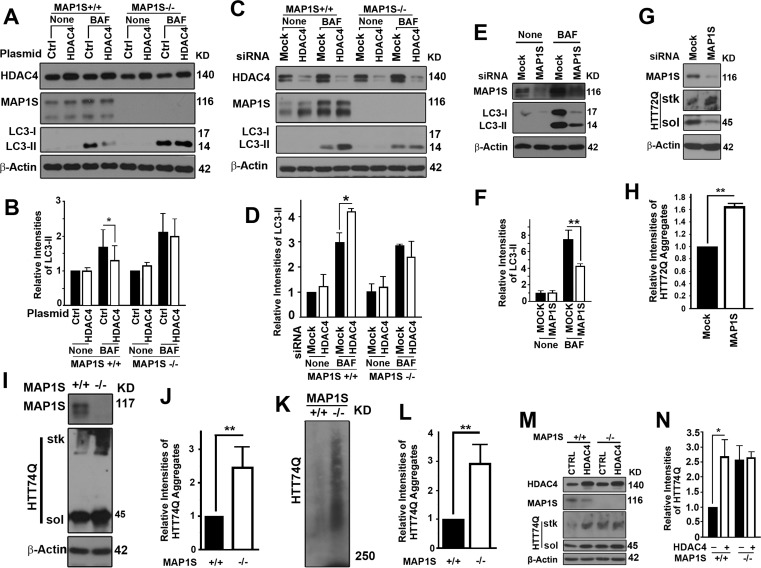
Figure 3.HDAC4 inhibits MAP1S-mediated autophagy clearance of mHTT aggregates(A-D) MAP1S is required for HDAC4 to affect LC3-II. Representative immunoblot results (A, C) and quantification (B, D) of the impact of MAP1S deletion on the effect of HDAC4 overexpression in wild-type and MAP1S−/− MEF cells (A, B) or suppression with siRNA in wild-type and MAP1S−/− HeLa cells (C, D) on LC3-II in the absence or presence of BAF. (E, F) MAP1S suppression with siRNA reduces levels of LC3-II in cells stably expressing HTT72Q. Representative immunoblots (E) and their quantification (F) are shown. (G, H) MAP1S suppression with siRNA increases levels of HTT72Q aggregates in cells stably expressing HTT72Q. Representative immunoblots (G) and their quantification (H) are shown. (I-L) MAP1S depletion increases levels of HTT74Q aggregates in MEF cells transiently expressing GFP-HTT74Q (HTT74Q). Representative immunoblot (I, K) and quantification (J, L) of the levels of HTT74Q aggregates in wild-type and MAP1S−/− MEF cells analyzed by stack gel (I, J) or AGERA (K, L). (M, N) MAP1S is required for the HDAC4-dependent increases in levels of HTT74Q aggregates. Representative immunoblots (M) and quantification (N) of the differences between wild-type and MAP1S−/− MEF cells are shown.